

Authors Mohan Kumar M et al
Cureus 18(10): e118479. doi:10.7759/cureus.118479
Abstract
Cerebral fat embolism syndrome (CFES) is a subset of fat embolism syndrome (FES) in which neurological manifestations predominate, making diagnosis using standard clinical criteria challenging. Large-vessel occlusion (LVO) associated with CFES is rare, with poor outcomes reported in the limited available literature. We present the case of a 74-year-old woman who sustained sequential bilateral femoral neck fractures following two separate mechanical falls. Both fractures were treated with hemiarthroplasties approximately 48 hours apart. Following the second operation, she failed to regain consciousness after general anaesthesia. CT angiography demonstrated occlusion in the right internal carotid artery (ICA) terminus extending into the M2 segment of the middle cerebral artery (MCA) with an additional distal M3 embolus. Subsequent MRI demonstrated a multi-territory starfield pattern alongside an established right MCA territorial infarct. Mechanical embolectomy was considered but not pursued because of the anticipated substantial postprocedural morbidity. Despite maximal supportive care, her neurological status remained unchanged. Given her poor overall prognosis, she was transitioned to comfort care and passed away. This case report highlights the diagnostic challenge of CFES without prominent respiratory or cutaneous manifestations, the value of early neuroimaging, and the emerging role of embolectomy in the LVO subtype. The closely spaced fractures and procedures raise the possibility of cumulative embolic exposure, although a causal relationship cannot be established from this case.
Introduction
Fat embolism syndrome (FES) is a recognised complication of fractures in which disseminated fat emboli in the systemic microvasculature cause endothelial injury. It typically follows long-bone trauma and manifests within 24-48 hours after injury, with a classic triad of respiratory failure, cutaneous petechiae, and neurological dysfunction [1]. Reported incidence estimates range from less than 1% to nearly 30%, with higher rates reported after bilateral femoral fractures [2,3]. This wide range reflects the syndrome’s nonspecific presentation, which limits consistent diagnosis. FES is primarily a clinical diagnosis supported by imaging findings and clinical scoring tools, such as the Gurd and Wilson, Lindeque, and Schonfeld criteria [2]. However, these clinical criteria have not been validated in prospective studies and lack well-established estimates of sensitivity or specificity [4].
Cerebral fat embolism syndrome (CFES) is a form of FES in which neurological manifestations predominate [5]. These range from mild confusion to focal deficits, seizures, and coma [6]. Failure to fulfil the classical diagnostic criteria, particularly in the absence of respiratory compromise, can make early recognition challenging [7]. Mild, nonspecific neurological symptoms may be overlooked without a high index of suspicion [4]. The true incidence of CFES is therefore difficult to establish. One prospective study identified magnetic resonance imaging (MRI) evidence of cerebral fat embolism in approximately 5% of a selected cohort of patients with long-bone or pelvic fractures [4].
Large-vessel occlusion (LVO) associated with CFES is rare. Poor outcomes have been described, although the available evidence is limited to selected published cases. Ooi et al. reported a series of three patients with cerebral LVO caused by fat embolism [8]. We present a fatal case of CFES with diffuse microemboli complicated by a coexisting proximal LVO. This case report highlights the diagnostic challenge of CFES without prominent respiratory or cutaneous manifestations, the value of early neuroimaging, and the emerging role of embolectomy in patients with the LVO subtype. The closely spaced fractures and procedures raise the possibility of cumulative embolic exposure, although a causal relationship cannot be established based on this case alone.
Case Presentation
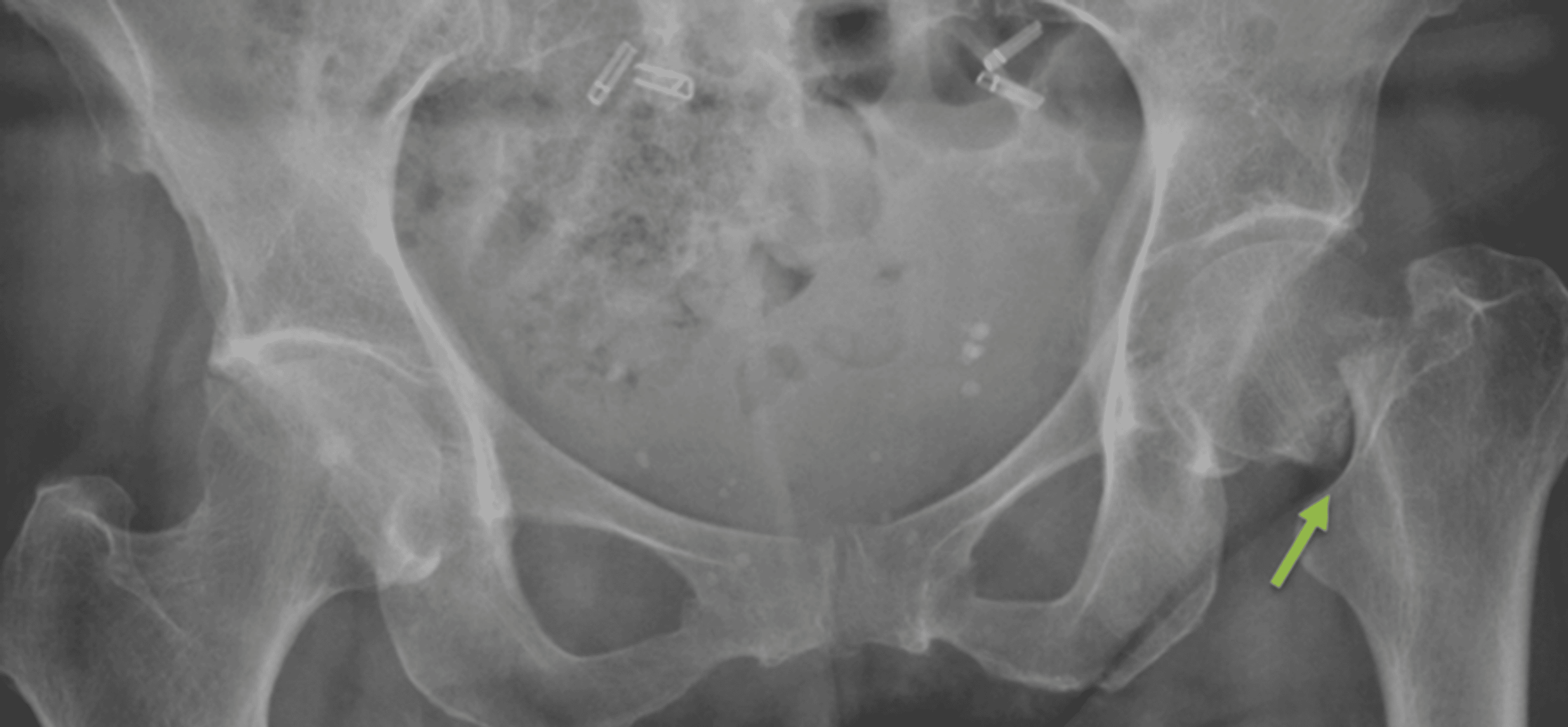
A 74-year-old, functionally independent woman presented to the emergency department after a mechanical fall onto her left hip without a head strike. Her medical history included stable triple-vessel ischaemic heart disease treated with percutaneous coronary intervention and coronary artery bypass grafting, breast cancer in remission, and hypothyroidism treated with replacement therapy. Plain radiography demonstrated a displaced left subcapital femoral neck fracture (Figure 1). She underwent left cemented hemiarthroplasty within 26 hours of the initial injury. The femoral canal was reamed, and a cement restrictor was placed; however, the use of pressurisation during cementing was not documented. General anaesthesia was administered without complication, and no intraoperative respiratory compromise was observed. She returned to the ward in a stable condition.
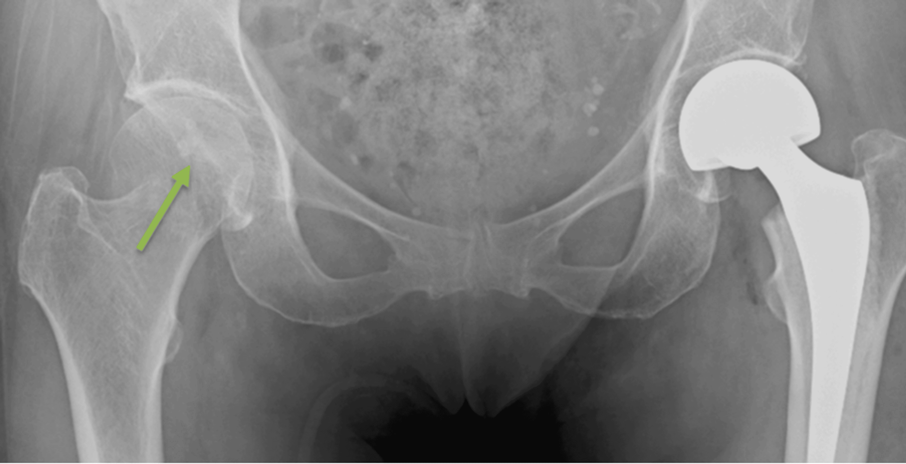
On the first postoperative day, the patient developed a mild oxygen requirement of 1 L/min via nasal prongs. Respiratory examination was normal, with no documented alteration in consciousness, petechiae, fever, or tachycardia. Haemoglobin concentration and platelet count remained stable. She was cleared for partial weight-bearing and mobilisation with a rollator under physiotherapy supervision. While mobilising to the bathroom on the ward 36 hours after surgery, she sustained an unwitnessed fall with a head strike. The fall was considered mechanical, and she remained neurologically intact following the event. Plain radiography identified an acute right subcapital femoral neck fracture (Figure 2); chest radiography was unremarkable. Non-contrast CT of the head showed periventricular deep white matter hypodensities consistent with microangiopathy, with no acute intracranial pathology identified. Given her persistent oxygen requirement, CT pulmonary angiography was performed and showed bilateral basal atelectasis, without pulmonary thromboembolism, consolidation, or ground-glass opacities.
Twelve hours after the second fall, the patient underwent right cemented hemiarthroplasty. The femoral canal was reamed, and pressurisation was not used during cementing. During cementing, she developed a brief episode of hypotension, with a systolic blood pressure of 50 mmHg, which initially did not respond to a 1 mg bolus of metaraminol. A further 0.5 mg bolus was followed by transient hypertension, with a systolic blood pressure of 200 mmHg, which was managed with propofol and sevoflurane. No hypoxia accompanied the hypotension, and end-tidal carbon dioxide remained stable.
She remained haemodynamically stable for the remainder of the operation without vasopressor support. There was no respiratory decompensation, persistent tachycardia or fever. Following cessation of anaesthesia and administration of reversal agents, she failed to awaken as expected. Intravenous naloxone, totalling 400 micrograms, produced no response. Her pupils remained equal and reactive, and no other focal neurological signs were identified at that time. She was triggering the ventilator with appropriate tidal volumes but remained unresponsive, with a Glasgow Coma Scale (GCS) score recorded as 3T (E1 VT M1), for approximately two hours. An arterial blood gas sample obtained 30 minutes after cessation of anaesthesia showed neither hypoxaemia nor hypercapnia. Electrolytes and blood glucose were within normal ranges. CT of the brain, CT angiography and CT perfusion were performed to investigate the persistent depression of consciousness.
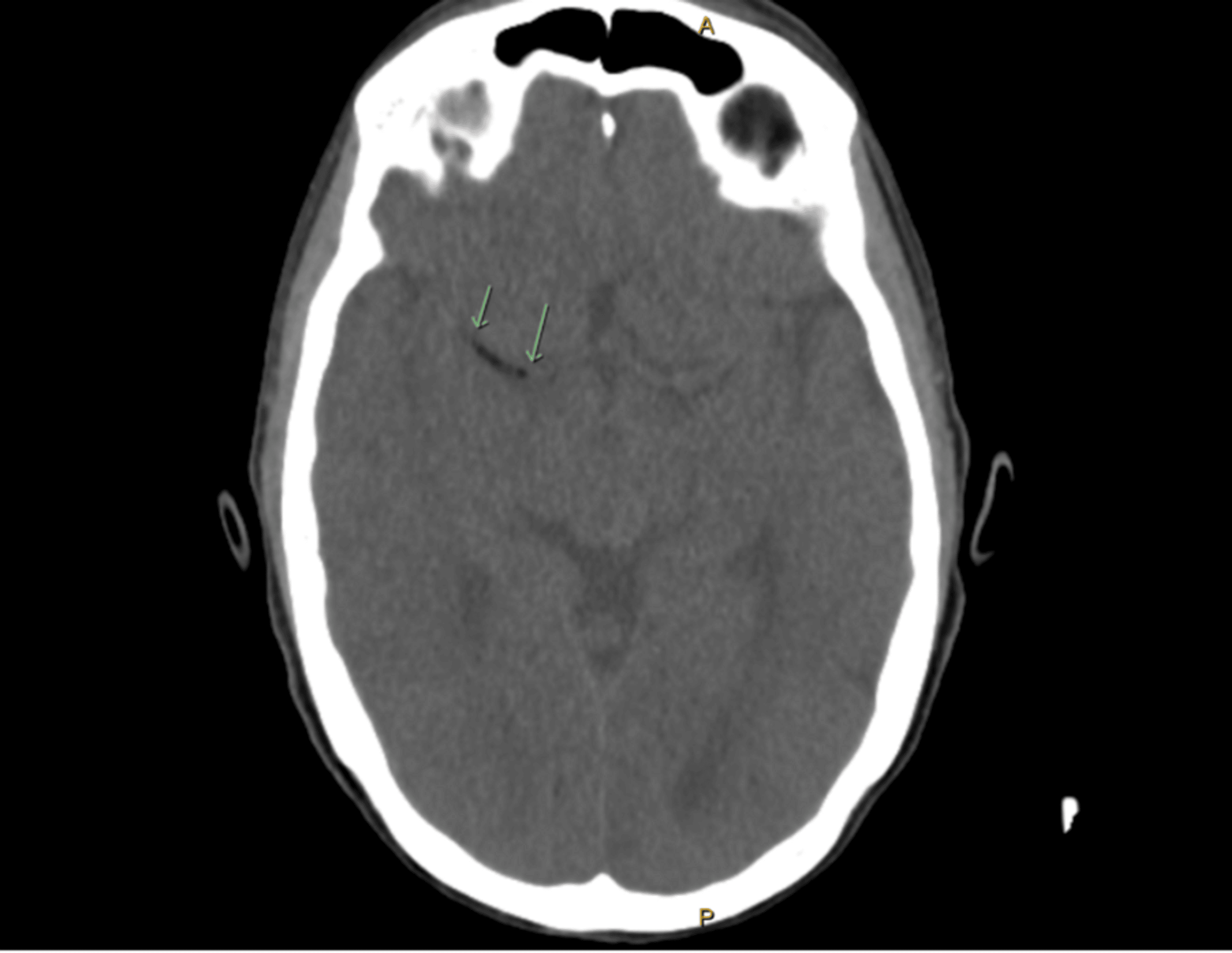
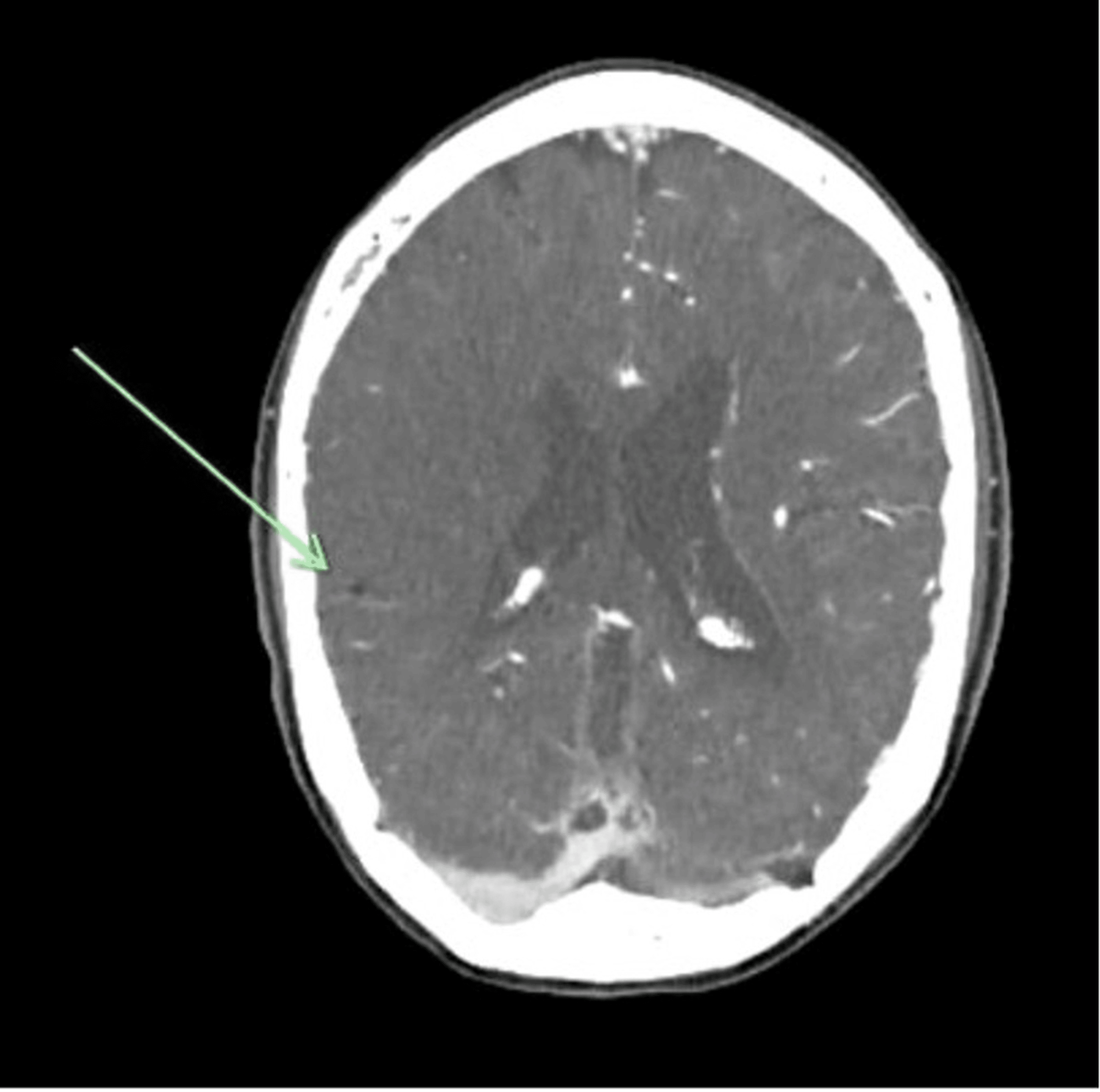
Non-contrast CT of the brain demonstrated early ischaemic changes in the right middle cerebral artery (MCA) territory with an intravascular hypodensity in the right M1 artery (Figure 3).
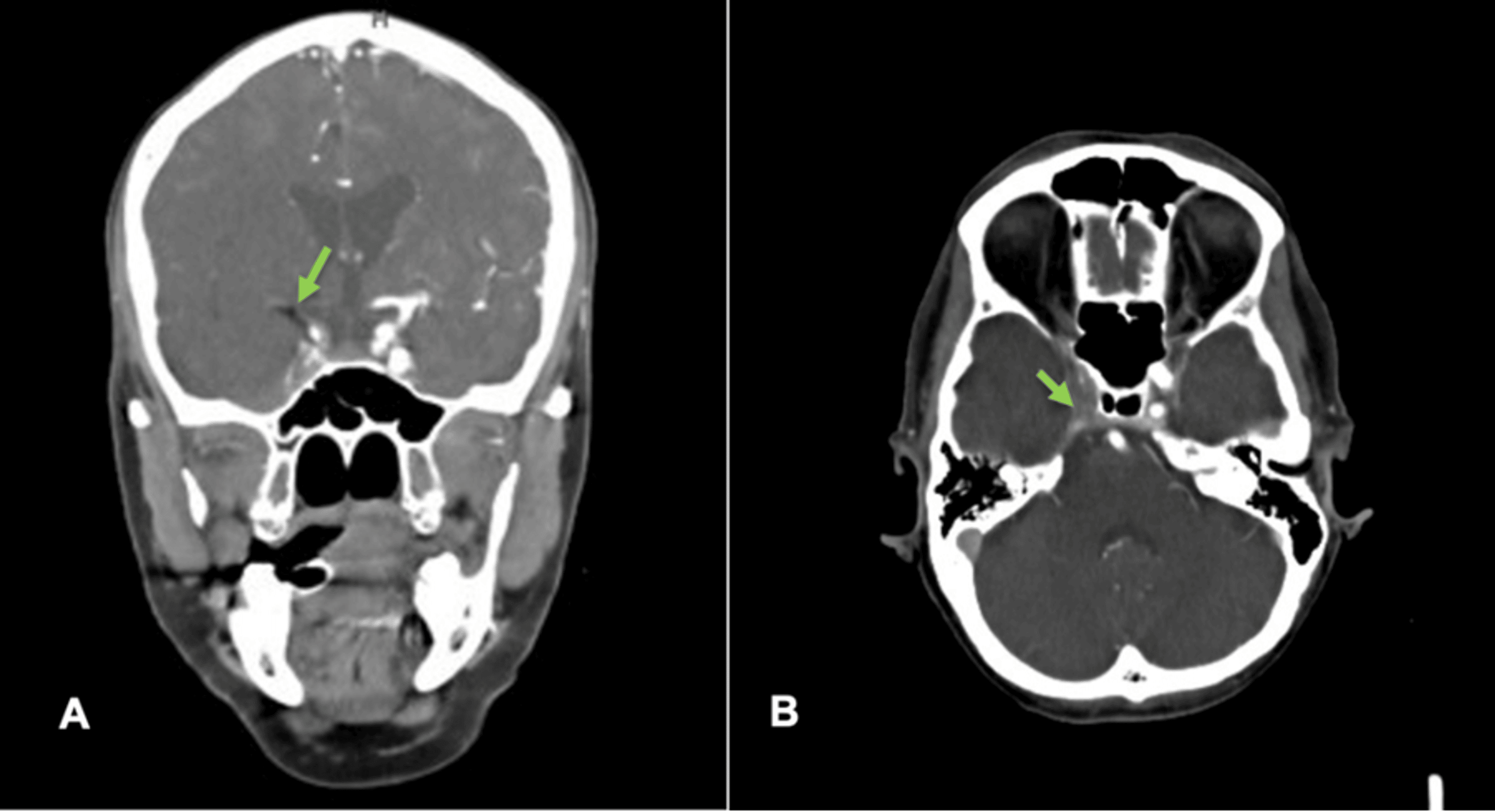
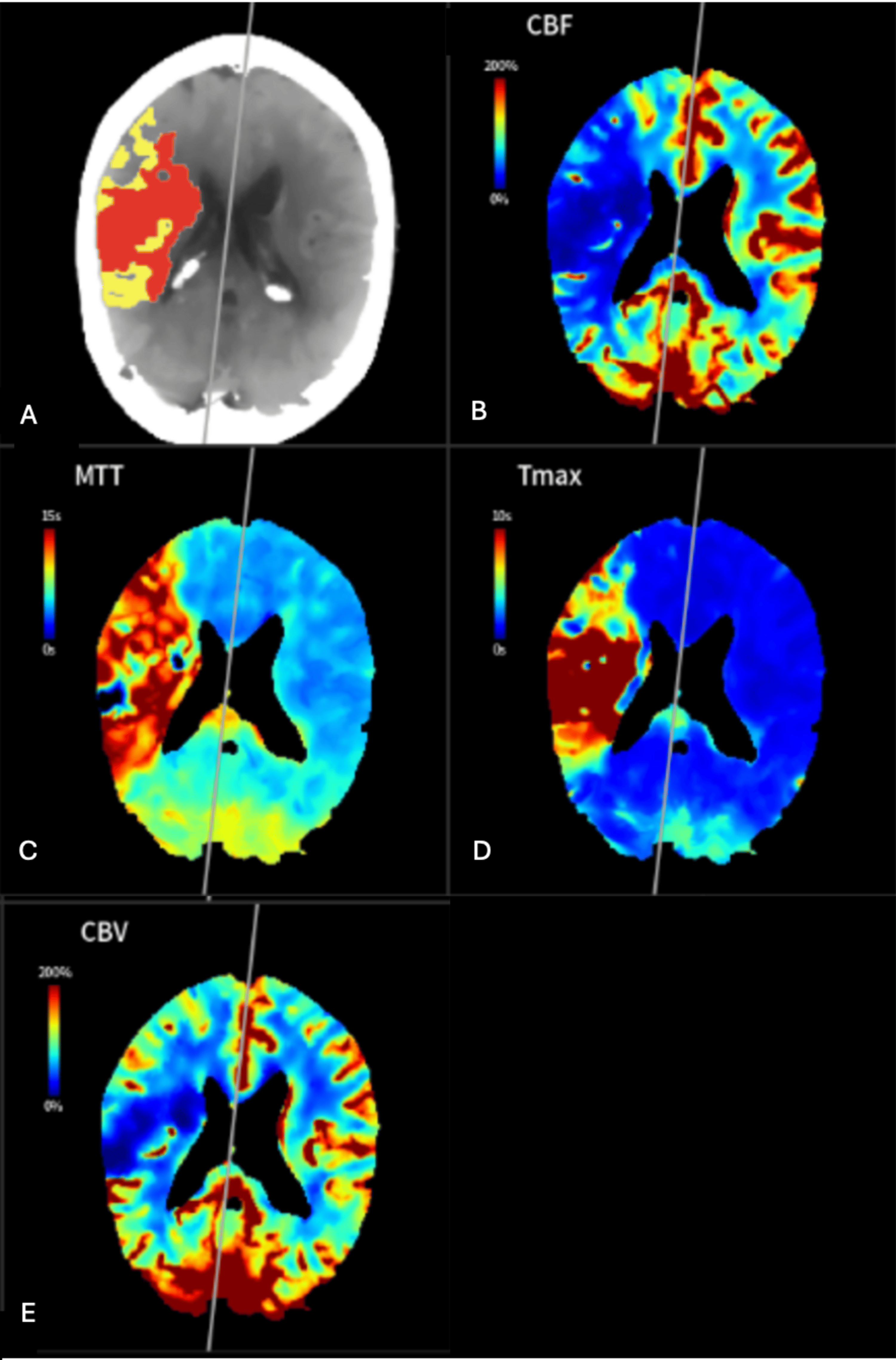
CT angiography demonstrated reduced opacification of the right internal carotid artery (ICA), attributed to downstream occlusion extending from the ICA terminus through the MCA to the M2 segment (Figure 4). Additionally, a distal embolus was identified in the M3 branch of the right MCA (Figure 5). CT perfusion estimated an ischaemic core of 123 mL and a penumbra of 34 mL (Figure 6). Based on the clinical course and initial imaging findings, a diagnosis of CFES was made at the time.
Mechanical embolectomy and intravenous thrombolysis were considered but not pursued; the rationale is explored in the discussion. The patient remained intubated, with a GCS score recorded as 3T (E1 VT M1), and was admitted to the ICU for neuroprotective and supportive care.
Levetiracetam was commenced approximately 18 hours after the onset of coma because of observed seizure activity. Neurosurgical consultation concluded that decompressive craniectomy would not be beneficial given the anticipated substantial postprocedural morbidity and mortality. Bedside transthoracic echocardiography did not demonstrate a patent foramen ovale (PFO); however, this was an informal study performed without agitated saline contrast. Repeat non-contrast CT of the brain approximately 48 hours after the onset of coma showed a large, established right MCA territorial infarct involving the right basal ganglia and caudate nucleus.
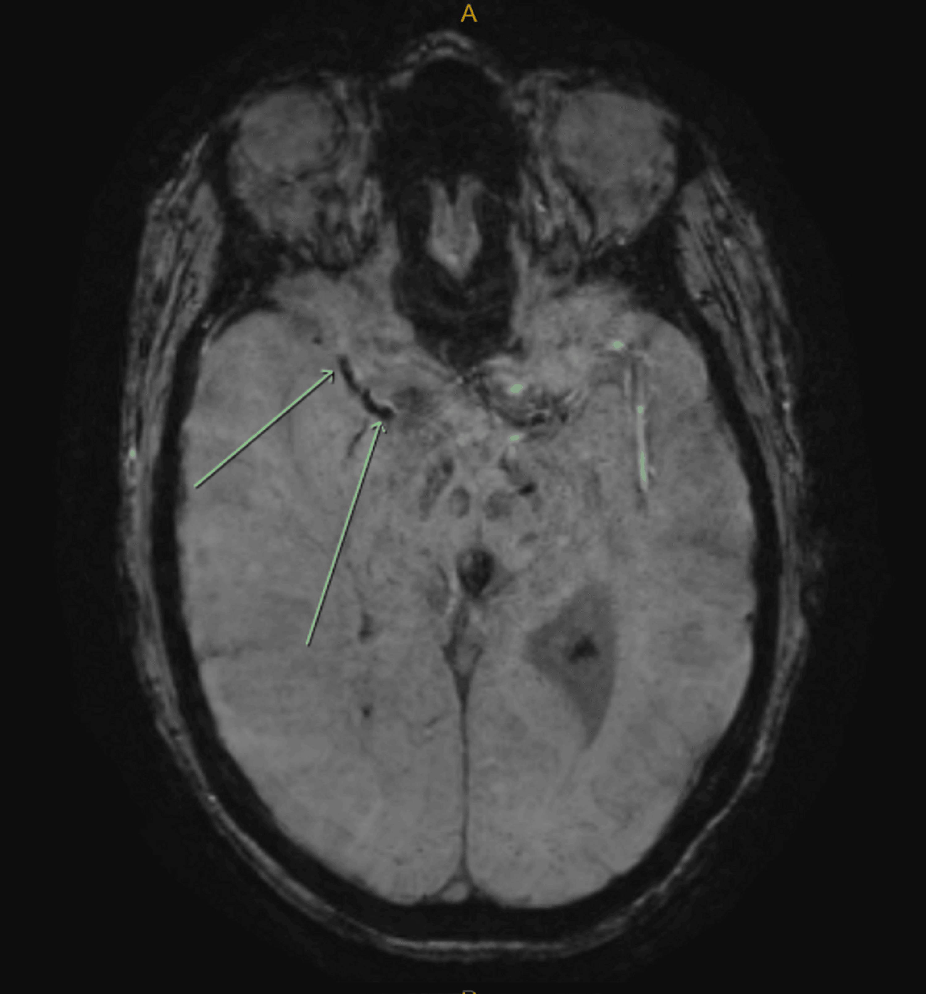
MRI was performed approximately 48 hours after the onset of coma to characterise the cerebral injury and assist with prognostic assessment. It demonstrated two coexisting patterns of injury: an extensive right MCA territorial infarct and numerous punctate lesions across multiple vascular territories. Susceptibility-weighted imaging (SWI) demonstrated an embolus in the right M1 artery (Figure 7). Time-of-flight imaging showed reduced flow in the petrous and cavernous segments of the right ICA and occlusion of the right M1 artery. Associated complications of the proximal LVO included substantial oedema, effacement of the lateral ventricle, and haemorrhagic transformation in the right basal ganglia.
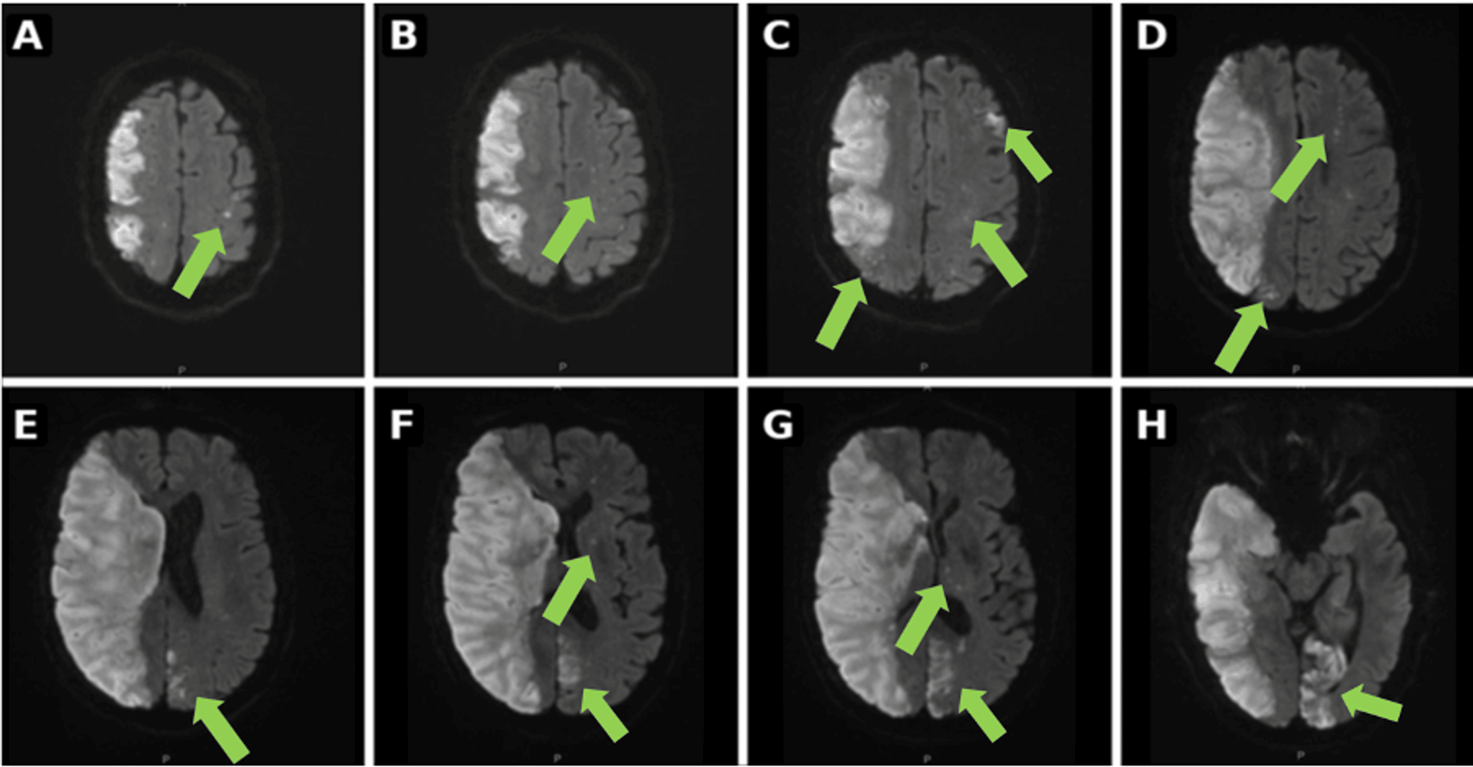
Diffusion-weighted imaging (DWI) demonstrated a multi-territory starfield pattern (Figure 8). Numerous punctate lesions were present in both anterior cerebral artery territories, the left basal ganglia and the right cerebellum, with particularly prominent involvement of the left posterior cerebral artery territory. DWI and the corresponding apparent diffusion coefficient (ADC) map demonstrated the extensive right MCA infarct alongside punctate left hemispheric lesions producing a starfield pattern (Figure 9).
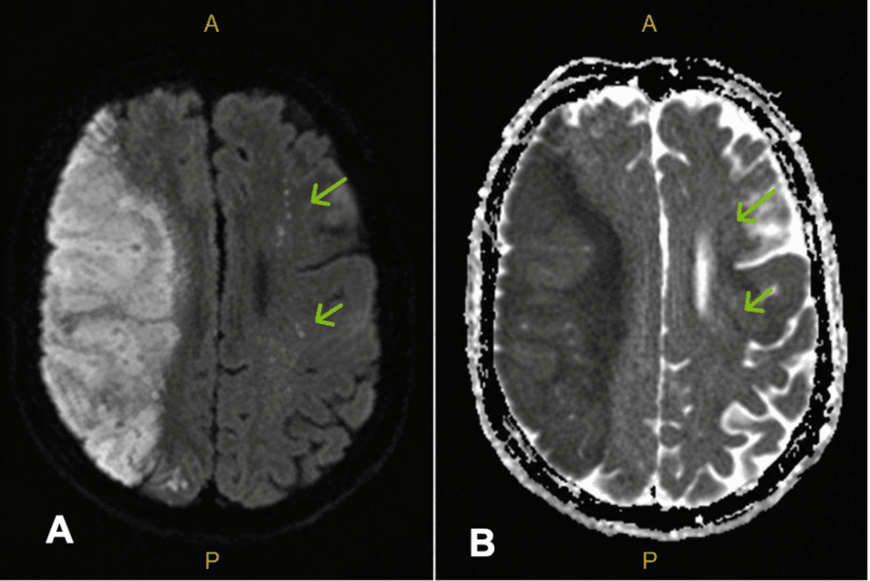
Serial neurological examinations showed no improvement, with the GCS score remaining recorded as 3T (E1 VT M1). The clinical and radiological findings indicated a poor overall prognosis, with substantial anticipated neurological morbidity. Following discussion with her next of kin, the patient was transitioned to comfort care and passed away. Her clinical course is summarised in Table 1.
Discussion
Mechanical and biochemical mechanisms have been proposed to explain FES. The mechanical theory suggests that raised intramedullary pressure forces marrow fat into the circulation, causing vascular obstruction and injury. The biochemical theory proposes catecholamine-driven inflammation and endothelial injury mediated by free fatty acids [1]. A PFO may facilitate paradoxical cerebral embolisation and has been implicated in CFES with LVO, although it is not a necessary precondition [8,9]. Fat has also been reported to traverse the pulmonary capillary bed directly, with catastrophic CFES described in the absence of a shunt identified at postmortem examination [10]. In this patient, bedside transthoracic echocardiography did not demonstrate a PFO, but the informal study was performed without agitated saline contrast and further investigations were not pursued. A shunt therefore could not be excluded. Both mechanisms, potentially aided by an undetected shunt, may have contributed to the coexistence of a proximal arterial embolus and diffuse cerebral microemboli.
The principal diagnostic challenge was the failure to emerge from anaesthesia without significant respiratory compromise. More common causes include residual sedation, bone cement implantation syndrome, postoperative encephalopathy and thromboembolic stroke. Fat-related arterial occlusion may be suggested on CT by low intravascular attenuation, in contrast to the hyperdense appearance of ordinary thrombus [9,11]. In this patient, non-contrast CT demonstrated an intravascular low-attenuation defect considered radiologically consistent with a fat embolus.
MRI, including DWI, SWI, T2-weighted sequences and gadolinium-enhanced imaging, can assist in diagnosing CFES. Imaging findings evolve through three described stages: an acute stage (days one to four), characterised by a scattered starfield pattern and early petechial haemorrhage; a subacute stage (days 5-14), marked by confluent white matter cytotoxic oedema; and a late stage (after day 14), in which resolving oedema may be followed by atrophy and gliosis. Petechial haemorrhage on SWI can persist across these stages and is considered a potentially characteristic finding; it was present in this case (Figure 10) [12].
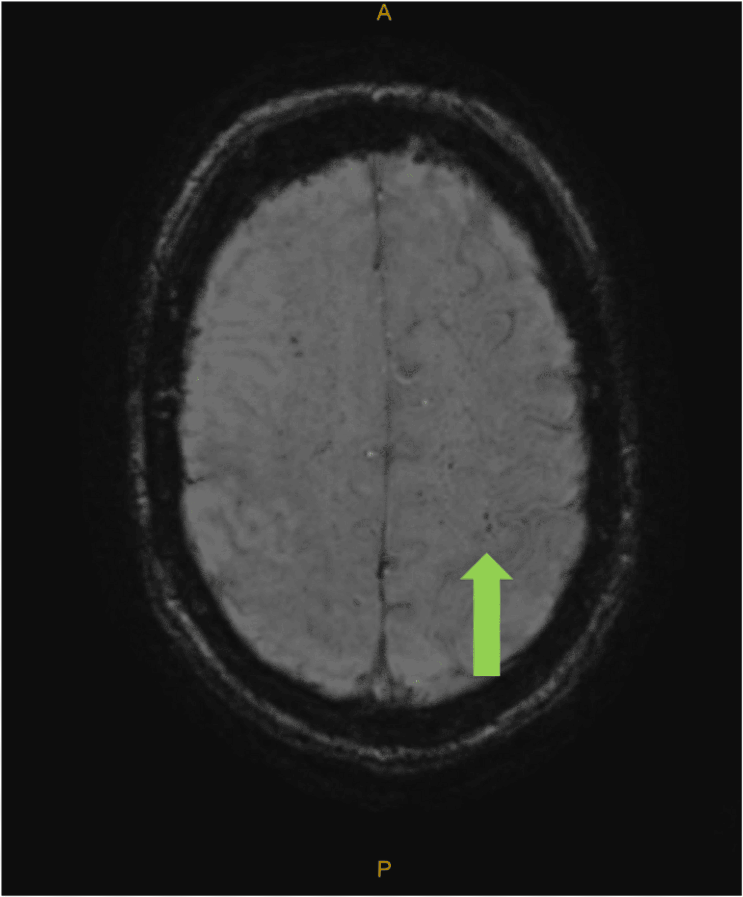
Imaging timing is important because a delayed scan may miss the acute pattern. In this patient, DWI demonstrated multiple punctate hyperintensities forming a starfield pattern, which has been proposed as an early, sensitive imaging marker of CFES [13]. The Takahashi MRI grading system assesses the size, burden, and distribution of lesions, primarily on T2-weighted sequences, and has been investigated as a prognostic tool. In a prospective cohort of 34 males with a mean age of 29.7 years, earlier symptom onset and higher MRI grade were associated with increased mortality [14]. These findings require cautious interpretation in an older woman with a concurrent major arterial occlusion.
MRI showed no evidence of brainstem infarction. Diffuse cerebral microembolic injury may have contributed to the depressed level of consciousness and was considered potentially reversible. However, serial neurological examinations over several days showed no improvement, with the GCS score remaining recorded as 3T (E1 VT M1). The principal concern was the extensive established right MCA infarction secondary to LVO, complicated by oedema and haemorrhagic transformation, which carried a high risk of severe neurological disability and death. Despite the potential reversibility of the diffuse microembolic injury, the combined clinical and radiological findings indicated a poor overall prognosis. Following discussion with her next of kin, the patient was transitioned to comfort care.
Mechanical embolectomy is increasingly reported as a diagnostic and therapeutic intervention for fat-related LVO. Retrieval permits direct visualisation and histopathological examination of the embolus [8,15]. Neither was possible in this patient because embolectomy was not performed. In a systematic review of 20 patients, Mudkavi et al. reported approximately 30% mortality following endovascular intervention. Mortality was lower with aspiration alone than with combined aspiration and stent retrieval, although the difference was not statistically significant [15]. These descriptive findings do not establish the superiority of either technique. Persistent microemboli that cannot be retrieved may contribute to incomplete recovery despite successful removal of a proximal embolus. In this patient, mechanical embolectomy was considered for the proximal LVO but was not pursued because of the anticipated substantial postprocedural morbidity and the assessment of her overall best interests, which were discussed with her next of kin. CT angiography also showed involvement of additional vascular territories, including the posterior circulation, and a distal M3 embolus that was not amenable to retrieval. Intravenous thrombolysis was considered but not pursued because she had recently undergone two major operations and the embolus was thought to be fat rather than thrombotic.
The timing of the second operation added complexity to this case. Two separate fractures and two definitive procedures in close succession could have increased cumulative embolic exposure, although this remains unproven. Historically, concern that early fixation could precipitate FES led to delayed approaches [3]. Delayed fixation has subsequently been associated with an increased risk of FES [2,3], and contemporary management generally favours stabilisation of long-bone fractures within 24-72 hours [3]. Multiple, displaced and bilateral femoral fractures are recognised risk factors [2,3]. Delaying treatment of the second fracture could also have prolonged pain and immobility. This case should therefore not be interpreted as evidence against early surgical treatment. In other fracture settings, techniques intended to reduce intramedullary pressure include external fixation, selected alternatives to intramedullary nailing, venting and reamer-irrigator-aspirator systems [2,3]. These approaches are not directly interchangeable with hemiarthroplasty for displaced femoral neck fractures.
Supportive care remains the mainstay of CFES management, including respiratory support, monitoring for raised intracranial pressure and treatment of seizures [6]. Other measures include optimising hydration, haemodynamics and nutrition, together with thromboembolism prophylaxis [2]. Prophylactic corticosteroids may reduce the incidence of FES, but this evidence should be distinguished from treatment of established CFES [2,16]. Corticosteroids are not recommended as standard treatment [6]. The patient received 4 mg of intravenous dexamethasone during the first operation and 8 mg during the second operation for anti-emesis as per the institution’s standard anaesthetic practice rather than for CFES prophylaxis. Transcranial Doppler ultrasonography can detect microembolic signals and may assist with monitoring high-risk patients [17]. Early hyperbaric oxygen therapy has been reported to improve neurological outcomes, but large-scale studies are lacking, and access may be limited [18]. Diffuse cerebral microembolic injury can be reversible, and good functional recovery has been reported among survivors [14]; nevertheless, overall mortality in CFES has been reported at approximately 10% [18]. These observations should not be directly extrapolated to patients with concurrent LVO and established territorial infarction.
This report has several limitations. It describes a single case retrospectively, and no causal inference can be made regarding the contribution of the fracture sequence or timing of surgery. Formal contrast echocardiography was not performed, and the use of pressurisation during the first cemented hemiarthroplasty was not documented. The diagnosis of fat embolism was based on the clinical course and imaging findings, without histopathological confirmation.
Conclusions
CFES with LVO is a rare but important differential diagnosis in trauma patients presenting with acute neurological decline and should be considered even in the absence of the classical respiratory and cutaneous manifestations of FES. Prompt neurological assessment, urgent vascular imaging when LVO is suspected, and timely MRI to provide additional diagnostic information are important for diagnosis and management. This case report illustrates the coexistence of these two patterns of injury following sequential bilateral femoral neck fractures and closely spaced operations. Although cumulative embolic exposure is plausible, a causal relationship cannot be established from a single case, and the outcome should not be interpreted as evidence in support of delayed surgical treatment. Vigilant postoperative monitoring and fall prevention remain important aspects of care.
References
- Godoy DA, Di Napoli M, Rabinstein AA: Cerebral fat embolism: recognition, complications, and prognosis. Neurocrit Care. 2018, 29:358-65. 10.1007/s12028-017-0463-y
- Shaikh N, Alali B, Amara UE, Nashrah UE, Alkheamy N, Ummunnisa F, Ghouri S: Fat embolism syndrome: evolving perspectives on diagnosis and care. Cureus. 2025, 17:e96136. 10.7759/cureus.96136
- Lempert M, Halvachizadeh S, Ellanti P, Pfeifer R, Hax J, Jensen KO, Pape HC: Incidence of fat embolism syndrome in femur fractures and its associated risk factors over time—a systematic review. J Clin Med. 2021, 10:14-6. 10.3390/jcm10122733
- Kanda N, Miyake T, Okada H, et al.: Prospective study examining the probability of cerebral fat embolism based on magnetic resonance imaging. Heliyon. 2023, 9:e14073. 10.1016/j.heliyon.2023.e14073
- Weinhouse GL: Fat embolism syndrome. UpToDate. 2026, 4:23-5.
- Morales-Vidal SG: Neurologic complications of fat embolism syndrome. Curr Neurol Neurosci Rep. 2019, 19:14. 10.1007/s11910-019-0928-9
- Lee SC, Yoon JY, Nam CH, Kim TK, Jung KA, Lee DW: Cerebral fat embolism syndrome after simultaneous bilateral total knee arthroplasty: a case series. J Arthroplasty. 2012, 27:409-14. 10.1016/j.arth.2011.06.013
- Ooi S, Raviskanthan S, Campbell BC, Hutton EJ, Mitchell PJ, Cloud GC: Cerebral large vessel occlusion caused by fat embolism—a case series and review of the literature. Front Neurol. 2021, 12:746099. 10.3389/fneur.2021.746099
- Maïer B, Badat N, Boulouis G, Zuber M: Mechanical thrombectomy for a cerebral fat embolism. Intensive Care Med. 2019, 45:1151. 10.1007/s00134-019-05518-z
- Berlot G, Bussani R, Shafiei V, Zarrillo N: Fulminant cerebral fat embolism: case description and review of the literature. Case Rep Crit Care. 2018, 2018:7813175. 10.1155/2018/7813175
- Alrasheed AS, Elazrag AM, Alqahtani MS, Alabbas F: Pathophysiology, clinical manifestations, and prognostic insights of cerebral fat embolism: a literature review. Front Neurol. 2025, 16:1732428. 10.3389/fneur.2025.1732428
- Kuo KH, Pan YJ, Lai YJ, Cheung WK, Chang FC, Jarosz J: Dynamic MR imaging patterns of cerebral fat embolism: a systematic review with illustrative cases. AJNR Am J Neuroradiol. 2014, 35:1052-7. 10.3174/ajnr.A3605
- Huang GS, Dunham CM, Chance EA: Occurrence of numerous cerebral white matter hyperintensities in trauma patients with cerebral fat embolism: a systematic review and report of two cases. Cureus. 2023, 15:e45450. 10.7759/cureus.45450
- Armstrong BR, Devendra A, Pokale S, et al.: Can the rate of mortality and neurological recovery be predicted from the time of onset of symptoms and MRI grade in patients with cerebral fat embolism? A study of 34 patients. Bone Joint J. 2022, 104-B:142-9. 10.1302/0301-620X.104B1.BJJ-2021-0420.R1
- Mudkavi R, Sarnecki T, Phyu EE, et al.: Cerebral fat embolism treated with mechanical embolectomy: case report and systematic review of the literature. BJR Case Rep. 2026, 12:uaag019. 10.1093/bjrcr/uaag019
- Schonfeld SA, Ploysongsang Y, DiLisio R, Crissman JD, Miller E, Hammerschmidt DE, Jacob HS: Fat embolism prophylaxis with corticosteroids. A prospective study in high-risk patients. Ann Intern Med. 1983, 99:438-43. 10.7326/0003-4819-99-4-438
- Jude M, Martin R, Nguyen I, et al.: Cerebral fat emboli monitoring using transcranial Doppler ultrasound and confirmation of a successful treatment response: a case report. J Trauma Inj. 2025, 38:152-8. 10.20408/jti.2024.0075
- Scarpino M, Lanzo G, Lolli F, Grippo A: From the diagnosis to the therapeutic management: cerebral fat embolism, a clinical challenge. Int J Gen Med. 2019, 12:39-48.