

Authors Errachki M et al
Cureus 18(10): e118061. doi:10.7759/cureus.118061
Abstract
Pneumocephalus, the accumulation of air within the intracranial compartments, most commonly follows head trauma with a skull-base or paranasal sinus fracture. When air continues to accumulate through a persistent osteodural defect and produces mass effect with clinical deterioration, it becomes tension pneumocephalus, a neurosurgical emergency.
We report the case of an 88-year-old man with a history of asthma who fell from standing height with a craniofacial impact during a three-day febrile gastrointestinal illness. After regaining consciousness at home, he developed progressive impairment of consciousness. The initial computed tomography (CT) scan, performed 48 hours after the fall, showed pneumocephalus with a basifrontal fracture involving the right orbital roof and lamina papyracea, in the setting of moderate traumatic brain injury (Glasgow Coma Scale (GCS) 10/15). Over the following 24 hours, his GCS decreased to 8/15, and repeat CT demonstrated tension pneumocephalus with a 9-mm midline shift and subfalcine herniation. Urgent frontal burr-hole decompression with subdural drainage achieved radiological improvement without recurrence.
Neurological recovery did not follow. The patient remained comatose (GCS 7/15) with a new severe left-sided hemiparesis. His intensive care course was complicated by an asthma exacerbation with dynamic hyperinflation, KDIGO stage 3 acute kidney injury, and a cardiac arrest of uncertain mechanism. He required a tracheostomy and died in the intensive care unit on day 31.
This case shows that established traumatic pneumocephalus may progress to tension pneumocephalus after a low-energy mechanism, and that any neurological decline in a patient with a skull-base fracture should prompt immediate repeat CT. Radiological success of decompression did not translate into neurological recovery, underscoring the weight of associated traumatic lesions and secondary systemic insults in elderly patients.
Introduction
Pneumocephalus is the presence of intracranial air and most often follows head trauma with a skull-base or paranasal sinus fracture [1]. Simple pneumocephalus produces no significant mass effect and usually resolves with conservative management. Tension pneumocephalus develops when air continues to accumulate through a persistent osteodural defect, producing mass effect with clinical deterioration, and requires urgent decompression [2]. The transition between these entities may be gradual and is recognized only through repeated clinical and radiological assessment [3,4].
We report the case of an 88-year-old man with moderate traumatic brain injury after a low-energy fall, in whom computed tomography (CT)-confirmed pneumocephalus associated with a basifrontal fracture progressed to tension pneumocephalus within 24 hours. The central learning point is that established traumatic pneumocephalus, even after a low-energy mechanism, may progress to tension and warrants close neurological monitoring with a low threshold for repeat imaging. The case also highlights the implications of a persistent osteodural defect for intensive care management, and the dissociation between radiological and neurological outcome.
Case Presentation
History and initial presentation
An 88-year-old man with a history of asthma (treatment regimen undocumented) developed a three-day illness with abdominal pain, diarrhea, and unmeasured fever suggestive of acute gastroenteritis. It was followed by acute muscular weakness, which resulted in a fall from standing height with a direct craniofacial impact. Witnesses reported a brief loss of consciousness and episodes of vomiting. The patient then regained consciousness and remained at home, but his condition subsequently worsened with progressive impairment of consciousness, which prompted medical consultation.
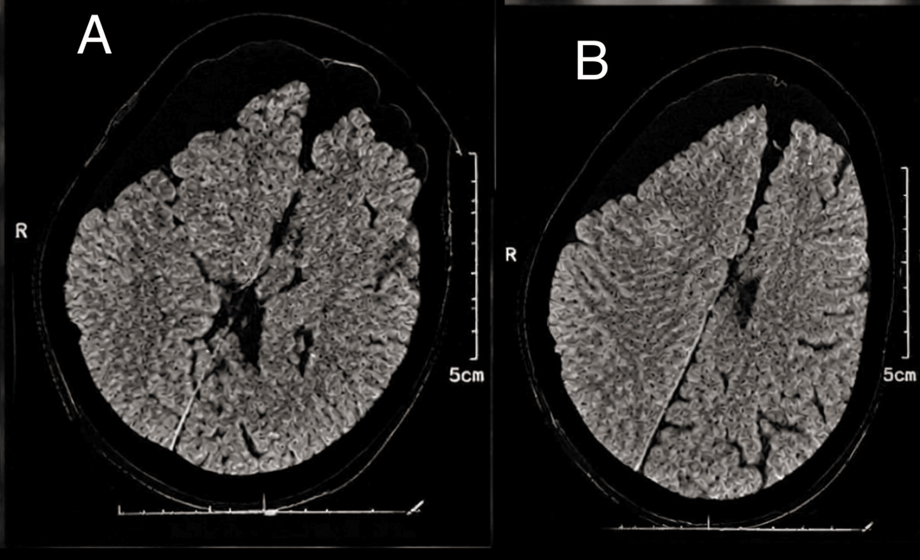
On arrival at the referring hospital, 48 hours after the fall, his Glasgow Coma Scale (GCS) was 10/15 (E2V3M5), consistent with moderate traumatic brain injury. Pupils were equal and reactive to light, and there was no focal motor deficit. There was no periorbital ecchymosis or other external sign of skull-base fracture, and no cerebrospinal fluid (CSF) rhinorrhea or otorrhea. The initial non-contrast brain CT (Figure 1) showed pneumocephalus predominantly involving the right frontoparietal region, associated with a basifrontal fracture of the right orbital roof and right lamina papyracea. Over the following hours, his GCS decreased to 8/15 (E1V2M5). A repeat CT performed approximately 24 hours after the initial scan (72 hours after the fall) showed progression of the pneumocephalus, and the patient was transferred to our tertiary hospital.
Admission to our emergency department
On arrival, the temperature was 38.5°C. The GCS was 8/15 (E1V2M5), with bilateral pinpoint pupils (reactivity; any sedative or opioid given during transfer) and spontaneous, symmetric movement of all four limbs without a lateralizing deficit. There was no rhinorrhea or otorrhea. The respiratory rate was 28 breaths/min, with an oxygen saturation of 92% on room air and bilateral coarse breath sounds. Blood pressure was 150/80 mmHg and heart rate 90 beats/min. Electrocardiography showed a left anterior fascicular block with right bundle branch block, secondary repolarization abnormalities, and frequent atrial ectopic beats. Transthoracic echocardiography showed a nondilated left ventricle with preserved global and regional systolic function and a nondilated right ventricle with preserved function. Admission laboratory findings are shown in Table 1.
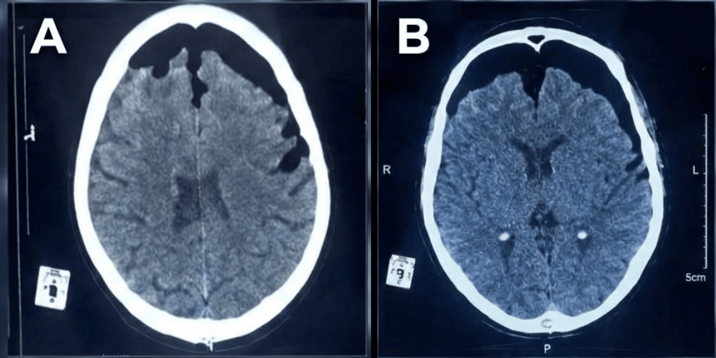
Emergency CT (Figure 2) showed tension pneumocephalus, with bilateral frontal subdural air compressing and separating the frontal lobes (Mount Fuji sign), a 9-mm leftward midline shift, and subfalcine herniation. Urgent surgical decompression was indicated.
Anesthesia and surgery
The patient was positioned supine with the head elevated to 30 degrees in a neutral position. Monitoring included continuous electrocardiography, noninvasive blood pressure, and pulse oximetry. After preoxygenation, rapid-sequence induction with cricoid pressure was performed; orotracheal intubation was uncomplicated (Cormack-Lehane grade I). Ceftriaxone 2 g was given for antibiotic prophylaxis. Anesthesia was maintained with target-controlled intravenous anesthesia combined with low-dose isoflurane, and nitrous oxide was avoided because of the intracranial air. A right femoral venous catheter and a right radial arterial catheter were inserted.
Through a 5-cm frontal incision, a frontal burr hole was created. After coagulation and opening of the dura mater, a Kehr drain was introduced into the subdural space and tunneled through a Redon drain. The procedure lasted approximately one hour, without major complication, transfusion, or vasopressor support. The anterior skull base was not explored, and the osteodural defect was not identified or repaired.
Intensive care course
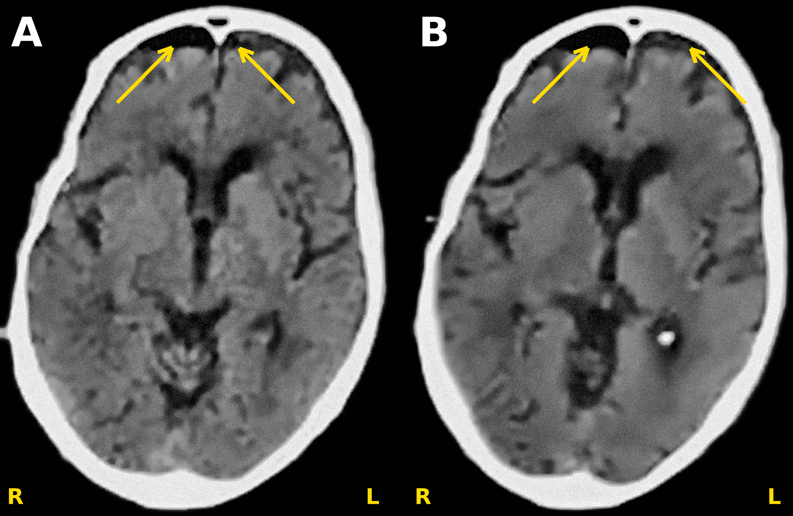
The patient was admitted to the surgical intensive care unit (ICU) sedated and mechanically ventilated. The subdural drain was connected to a closed gravity drainage system with a dependent collection bag and initially drained approximately 50 mL of serohematic fluid containing air bubbles. Management focused on preventing secondary brain insults (blood pressure, oxygenation, ventilation, temperature, glucose, and sodium) with serial neurological and pupillary assessments. Postoperative CT (Figure 3A) showed regression of the pneumocephalus, with (right/bilateral) subcortical and deep frontal white-matter contusions consistent with a coup-contrecoup injury and a spontaneous hyperdensity of the cerebellar vermis and hemispheres, suggesting possible traumatic subarachnoid hemorrhage. Sedation was then discontinued.
Approximately 12 hours after sedation withdrawal, the GCS was 7/15 and the Full Outline of UnResponsiveness (FOUR) score 9/16, with equal pupils reactive to light. A severe left-sided hemiparesis, graded 1/5 in both the upper and lower limbs and absent on preoperative examination, was observed; the right limbs remained mobile. No clinical seizures were observed, and an electroencephalogram showed no evidence of nonconvulsive status epilepticus.
On ICU day 3, the patient developed an asthma exacerbation with bilateral wheezing and dynamic hyperinflation; the expiratory flow curve did not return to baseline. Intratracheal salbutamol nebulization and adjustment of ventilator settings improved gas exchange.
On day 5, the subdural drain was removed after air and fluid output had ceased. To limit recurrent air entry, the head of the bed was kept at 30 degrees, nasal suctioning and nasogastric tubes were avoided, and airway pressures were limited. No CSF rhinorrhea or otorrhea was observed at any time during the ICU stay.
Also on day 5, the patient presented with new neurological deterioration, together with persistent fever and an inflammatory response without an identified source (C-reactive protein 157 mg/L). Because of the skull-base fracture, post-traumatic meningitis had to be excluded. A brain CT performed beforehand showed no recurrence of the pneumocephalus, no midline shift, patent basal cisterns, and no hydrocephalus or posterior fossa mass effect. A lumbar puncture was then performed. The CSF was hemorrhagic, with a protein level of 6.8 g/L, a low leukocyte count, and normal glucose. CSF, protected distal airway, and urine cultures were negative.
The same day, acute kidney injury developed, initially KDIGO stage 1 and progressing to stage 3, with transient oliguria followed by recovery of urine output, metabolic acidosis, and hyperkalemia up to 6.53 mmol/L (Table 2). An urgent nephrology consultation was obtained. Renal replacement therapy was considered but not initiated, as urine output recovered and no emergency indication (refractory hyperkalemia, refractory metabolic acidosis, or fluid overload) developed.
On day 7, the patient developed progressive bradycardia to 40 beats/min, followed by asystole. Return of spontaneous circulation was achieved after approximately five minutes of cardiopulmonary resuscitation and epinephrine administration. At the time of the arrest, he was ventilated in volume-controlled mode with a tidal volume of 470 mL, a respiratory rate of 18 breaths/min, and an external positive end-expiratory pressure (PEEP) of 7 cmH₂O. The plateau pressure was 27 cmH₂O, and an end-expiratory occlusion revealed an intrinsic PEEP of 6 cmH₂O (total PEEP 13 cmH₂O). Arterial blood gas analysis obtained shortly before the arrest showed a pH of 7.27, a PaCO₂ of 50 mmHg, and a PaO₂ of 80 mmHg on an FiO₂ of 0.60 (PaO₂/FiO₂ ratio 133). Serum potassium was 5.2 mmol/L. The mechanism of the arrest was not established. Post-arrest care included optimization of oxygenation, maintenance of mean arterial pressure above 75 mmHg, and close metabolic monitoring.
A follow-up CT on day 10 (Figure 3B) showed slight further regression of residual frontal air bubbles without recurrence; the frontal contusions and posterior fossa hyperdensity were stable (with no new infarction or vascular territory abnormality). No MRI or vascular imaging was performed.
Outcome
Given persistent impaired consciousness and the anticipated need for prolonged mechanical ventilation, a tracheostomy was performed on day 12 to facilitate airway management and nursing care. Neurological status did not improve: at the last assessment, the GCS was 7/15 and the FOUR score 9/16, with persistent severe left-sided hemiparesis. Renal function did not recover (last serum creatinine 43 mg/L, approximately 380 µmol/L). The patient remained dependent on mechanical ventilation and died in the ICU on day 31. The main events are summarized in Table 2.
Discussion
Pneumocephalus may be traumatic, iatrogenic, infectious, or, rarely, spontaneous [4,5]. Traumatic pneumocephalus occurs in approximately 0.5% to 1% of head injuries, and progression to tension pneumocephalus is uncommon [6]; other recognized precipitants include neurosurgical and otorhinolaryngological procedures, positive-pressure ventilation, nitrous oxide anesthesia, and air travel [7]. The central observation in this case is the progression of established traumatic pneumocephalus to tension pneumocephalus after a low-energy mechanism. The case also illustrates the intensive care implications of a persistent osteodural defect and the dissociation between radiological and neurological outcome.
Progression of established pneumocephalus
In our patient, consciousness was initially preserved after the fall, and neurological impairment developed progressively over the following 48 hours. Pneumocephalus was already present on the initial CT and progressed to tension pneumocephalus, with a 9-mm midline shift and subfalcine herniation, over the next 24 hours. Tension may develop days after the initial injury as long as the osteodural defect persists [8], with a variable latency [9], and its most common manifestations, headache and altered consciousness, are nonspecific [10,11]. A low-energy mechanism therefore should not be reassuring when a skull-base fracture is present, and any decline in neurological status should prompt repeat imaging [3,4].
Mechanism
The subdural space offers little resistance to gas expansion and is a preferential site for tension pneumocephalus [12,13]. In our patient, progressive air entrapment through the skull-base defect, consistent with a ball-valve mechanism [14,15], is the most plausible explanation, but it was not directly demonstrated. Supporting findings include the basifrontal fracture involving the orbital roof and lamina papyracea, the predominantly frontal subdural collection, radiological enlargement over 24 hours, and repeated vomiting after the fall. The absence of clinical rhinorrhea makes an inverted-bottle mechanism less likely [15], although an occult CSF leak cannot be excluded.
Diagnosis and surgical management
Non-contrast CT can detect as little as 0.5 cm³ of intracranial air [16], and the Mount Fuji sign, present in our patient, is highly suggestive of tension [17]. Conservative management is appropriate for simple pneumocephalus, whereas neurological deterioration with mass effect requires urgent decompression [2,6,18]. Burr-hole evacuation with subdural drainage achieved rapid radiological improvement. Nitrous oxide was avoided because of its potential to expand intracranial air collections. Normobaric oxygen may accelerate air resorption but remains an adjunct rather than an alternative to decompression [8].
Radiological versus clinical outcome
Decompression was technically and radiologically successful, with regression of the pneumocephalus and no recurrence, but it was not followed by neurological recovery: the GCS remained 7/15 from sedation withdrawal until death, with a severe left-sided hemiparesis. Several factors may account for this dissociation. First, associated traumatic lesions were present, including frontal contusions consistent with a coup-contrecoup injury and a probable traumatic subarachnoid hemorrhage in the posterior fossa, supported by the hemorrhagic CSF. Second, the brain had been exposed to mass effect with subfalcine herniation for an undetermined period before decompression, and ischemic injury not detectable on CT cannot be excluded. The appearance of a left-sided hemiparesis after surgery, absent preoperatively and consistent with right-hemispheric injury on the side of the predominant air collection and mass effect, supports this hypothesis. Third, the patient sustained repeated secondary systemic insults, including fever, impaired oxygenation, hypercapnic acidosis, electrolyte disturbances, acute kidney injury, and a cardiac arrest; the arrest occurred after the deficits were first observed and did not cause them, but it may have compromised subsequent recovery. Advanced age likely further limited neurological reserve. Nonconvulsive status epilepticus was excluded by electroencephalography, but in the absence of MRI, the relative contribution of each factor could not be determined.
Intensive care considerations
The osteodural defect was not identified or repaired, so the risk of recurrent air entry persisted throughout the ICU stay. After drain removal on day 5, it was managed by head-of-bed elevation, avoidance of nasal suctioning and nasogastric tubes, and limitation of airway pressures, and follow-up CT showed no recurrence. Positive-pressure ventilation, including noninvasive modalities, may promote air entry when a communication with the airways persists [1,9,19], and this constraint became critical during the asthma exacerbation. At the time of the cardiac arrest, an intrinsic PEEP of 6 cmH₂O indicated persistent dynamic hyperinflation. Dynamic hyperinflation raises intrathoracic pressure, which reduces venous return and right ventricular preload and may cause hemodynamic collapse independently of hypoxemia. The bradycardia preceding asystole, the hypercapnic acidosis (pH 7.27, PaCO₂ 50 mmHg), and the impaired oxygenation (PaO₂/FiO₂ 133) without severe hypoxemia are compatible with a combined respiratory and hemodynamic mechanism. However, no ventilator disconnection test or advanced hemodynamic monitoring was documented before the arrest, and severe hyperkalemia was absent (potassium 5.2 mmol/L), so the mechanism remains uncertain.
Lumbar puncture is contraindicated in the presence of intracranial mass effect, and CSF withdrawal may favor air entry when an osteodural defect persists. In our patient, it was performed only on day 5, after decompression and after CT had excluded recurrent pneumocephalus and mass effect, because excluding meningitis was essential in a febrile patient with a skull-base fracture. The hemorrhagic CSF with elevated protein, a low leukocyte count, normal glucose, and negative cultures favored a post-traumatic rather than infectious origin. Awareness of these interactions by intensivists is essential for prompt recognition and management [20].
Conclusions
In this patient with moderate traumatic brain injury and a basifrontal skull-base fracture, established traumatic pneumocephalus progressed to tension pneumocephalus with midline shift and subfalcine herniation within 24 hours of the initial CT. When pneumocephalus is associated with a skull-base fracture, any decline in GCS or failure to improve should be regarded as a possible sign of developing intracranial mass effect and should prompt immediate repeat CT, regardless of the energy of the injury mechanism. Burr-hole decompression achieved rapid radiological improvement without recurrence, but it was not followed by neurological recovery: the patient remained comatose with a severe left-sided hemiparesis and died in the ICU on day 31 after a course complicated by secondary systemic insults. Radiological success of decompression should therefore not be equated with clinical recovery, particularly in elderly patients with associated traumatic lesions.
References
- Wankhade BS, Beniamein MM, Alrais ZF, Mathew JI, Alrais GZ: What should an intensivist know about pneumocephalus and tension pneumocephalus?. Acute Crit Care. 2023, 38:244-8. 10.4266/acc.2021.01102
- Schirmer CM, Heilman CB, Bhardwaj A: Pneumocephalus: case illustrations and review. Neurocrit Care. 2010, 13:152-8. 10.1007/s12028-010-9363-0
- Śmiłowska K, Sznajder-Stacha K, Kocyłowski D, Popek A, Rozpondek K, Grechuta M, Salari M: Pneumocephalus as a rare complication: a systematic review plus clinical vignette. Neurol Neurochir Pol. 2021, 55:253-68. 10.5603/PJNNS.a2021.0014
- Sarwal A: Neurologic complications in the postoperative neurosurgery patient. Continuum (Minneap Minn). 2021, 27:1382-404. 10.1212/CON.0000000000001039
- Horowitz M: Intracranial pneumatocele – an unusual complication following mastoid surgery. J Laryngol Otol. 1964, 78:128-34. 10.1017/s0022215100061910
- Pillai P, Sharma R, MacKenzie L, Reilly EF, Beery PR 2nd, Papadimos TJ, Stawicki SP: Traumatic tension pneumocephalus – two cases and comprehensive review of literature. Int J Crit Illn Inj Sci. 2017, 7:58-64. 10.4103/IJCIIS.IJCIIS_8_17
- Sweni S, Senthilkumaran S, Balamurugan N, Thirumalaikolundusubramanian P: Tension pneumocephalus: a case report with review of literature. Emerg Radiol. 2013, 20:573-8. 10.1007/s10140-013-1135-7
- Gore PA, Maan H, Chang S, Pitt AM, Spetzler RF, Nakaji P: Normobaric oxygen therapy strategies in the treatment of postcraniotomy pneumocephalus. J Neurosurg. 2008, 108:926-9. 10.3171/JNS/2008/108/5/0926
- Chang Y, Kim TG, Chung SY: High-flow nasal cannula-induced tension pneumocephalus. Indian J Crit Care Med. 2020, 24:592-5. 10.5005/jp-journals-10071-23482
- Garonzik IM, Samdani AF, Carson BS, Avellino AM: Pneumocephalus in a newborn with an open myelomeningocele. Pediatr Neurosurg. 2001, 35:334. 10.1159/000050447
- Harth C, Peeters I, Van Hoe L, Cypers G, Dekeyzer S: Intrathecal gadolinium-enhanced MR-cisternography for the detection of a petrous bone defect in a patient with spontaneous intraventricular pneumocephalus. Acta Neurol Belg. 2020, 120:695-8. 10.1007/s13760-019-01121-7
- Lunsford LD, Maroon JC, Sheptak PE, Albin MS: Subdural tension pneumocephalus. Report of two cases. J Neurosurg. 1979, 50:525-7. 10.3171/jns.1979.50.4.0525
- Harlyjoy A, Nathaniel M, Nugroho AW, Gunawan K: Traumatic tension pneumocephalus: a case report and perspective from Indonesia. Front Neurol. 2024, 15:1339521. 10.3389/fneur.2024.1339521
- Monas J, Peak DA: Spontaneous tension pneumocephalus resulting from a scalp fistula in a patient with a remotely placed ventriculoperitoneal shunt. Ann Emerg Med. 2010, 56:378-81. 10.1016/j.annemergmed.2010.05.030
- Ishiwata Y, Fujitsu K, Sekino T, Fujino H, Kubokura T, Tsubone K, Kuwabara T: Subdural tension pneumocephalus following surgery for chronic subdural hematoma. J Neurosurg. 1988, 68:58-61. 10.3171/jns.1988.68.1.0058
- Osborn AG, Daines JH, Wing SD, Anderson RE: Intracranial air on computerized tomography. J Neurosurg. 1978, 48:355-9. 10.3171/jns.1978.48.3.0355
- Agrawal A, Singh BR: Mount Fuji sign with concavo-convex appearance of epidural haematoma in a patient with tension pneumocephalus. J Radiol Case Rep. 2009, 3:10-2. 10.3941/jrcr.v3i1.100
- Arai N, Tabuse M, Nakamura A, Miyazaki H: Spontaneous intraventricular pneumocephalus. J Craniofac Surg. 2017, 28:738-40. 10.1097/SCS.0000000000003392
- Kopelovich JC, de la Garza GO, Greenlee JD, Graham SM, Udeh CI, O’Brien EK: Pneumocephalus with BiPAP use after transsphenoidal surgery. J Clin Anesth. 2012, 24:415-8. 10.1016/j.jclinane.2011.12.003
- Amato-Watkins A, Rao VM, Leach P: Air travel after intracranial surgery: a survey of advice given to patients by consultant neurosurgeons in the UK. Br J Neurosurg. 2013, 27:9-11.