

Authors Chahboun B, Majit A, Kaba O, et al.
Cureus 18(10): e118204. doi:10.7759/cureus.118204
Abstract
Hemorrhagic stroke is rare in children but carries a substantial risk of mortality and long-term neurological disability. Its presentation may be nonspecific, with seizures and impaired consciousness potentially mimicking infectious or metabolic disorders. We report the case of an eight-year-old girl with no personal or family history of neurological disease and reportedly normal psychomotor development who presented with seizures, vomiting, and impaired consciousness. The initial computed tomography report described a left parietal intraparenchymal hematoma associated with intraventricular hemorrhage and mass effect. Emergency surgical evacuation was performed under general anesthesia, with intraoperative osmotherapy and invasive hemodynamic monitoring. The postoperative course was characterized by progressive recovery of consciousness, although right hemiplegia and aphasia persisted. Brain magnetic resonance imaging performed on postoperative day three showed an evolving intraparenchymal hematoma without appreciable contrast enhancement. Three-dimensional time-of-flight magnetic resonance angiography demonstrated preserved patency of the major intracranial arteries of the circle of Willis, without an apparent intracranial aneurysm or significant arterial stenosis. However, the available investigations did not establish the cause of the hemorrhage, and cerebral venous thrombosis or an occult vascular lesion could not be excluded. Multidisciplinary rehabilitation was initiated early. At six months, the patient was independent in the main activities of daily living and walked without an assistive device, with residual mild right-sided motor impairment, reduced right-hand dexterity, and mild language difficulties. This case illustrates the importance of rapid multidisciplinary management and continued rehabilitation after pediatric intracerebral hemorrhage, as well as the need for a complete etiological evaluation and appropriate vascular follow-up when the initial investigations are inconclusive.
Introduction
Pediatric stroke is rare, with a recent systematic review estimating an overall childhood stroke incidence of approximately 3.73 per 100,000 person-years and a hemorrhagic stroke incidence of approximately 0.99 per 100,000 person-years [1]. Pediatric hemorrhagic stroke differs from adult intracerebral hemorrhage in its etiological spectrum, with vascular abnormalities, hematological disorders, systemic conditions, tumors, and infections among the recognized causes [2,3]. Vascular etiologies are particularly important in children, with arteriovenous malformations representing the most frequently reported vascular cause in systematic reviews [2].
The clinical presentation may be nonspecific and can include seizures, vomiting, headache, impaired consciousness, and, occasionally, fever. Such manifestations may initially suggest an infectious or metabolic disorder and potentially delay neuroimaging and definitive management [3,4]. Atypical presentations of intracranial hemorrhage may require evaluation for several possible diagnoses before neuroimaging establishes the underlying structural lesion. Prompt neuroimaging is therefore essential in children with unexplained acute neurological deterioration. The subsequent etiological work-up may require dedicated arterial and venous imaging, and digital subtraction angiography may be indicated when the initial vascular evaluation does not establish a cause [4].
We report the case of an eight-year-old girl who presented with seizures and impaired consciousness associated with an intracerebral hemorrhage with intraventricular extension. The case illustrates the acute anesthetic and intensive care management, the limitations of the available postoperative vascular assessment, and subsequent multidisciplinary rehabilitation over a six-month period. The learning objectives are to emphasize early recognition of pediatric intracerebral hemorrhage, the distinction between an initially negative noninvasive vascular study and a definitive etiological diagnosis, and the importance of functional follow-up after acute treatment.
Case Presentation
An eight-year-old girl with no personal or family history of neurological disease and reportedly normal psychomotor development was referred from a provincial hospital for management of impaired consciousness. The initial symptoms developed at home and consisted of seizures, vomiting, and a brief episode of hyperthermia that prompted the initial medical consultation. The child remained afebrile from the time of transfer until admission to our unit. The available documentation did not provide sufficient detail to establish the seizure type, individual seizure duration, or total seizure burden. At the referring facility, the Glasgow Coma Scale (GCS) score was 8/15, with a motor score of 4, verbal score of 2, and eye-opening score of 2. Blood pressure was 120/60 mmHg and heart rate was 80 beats/min, without prominent respiratory distress. Given the severe neurological impairment, rapid-sequence orotracheal intubation was performed with cricoid pressure after administration of fentanyl, propofol, and rocuronium, using a size 5 endotracheal tube. The patient remained hemodynamically stable during medically supervised transfer.
On admission to the pediatric intensive care unit, capillary blood glucose was 1.02 g/L (reference range: 0.70-1.10 g/L) and body temperature was 37.2°C. The conjunctivae were normally colored. The patient was deeply sedated, with no response to voice or physical stimulation. Blood pressure was 100/56 mmHg and heart rate was 90 beats/min. Oxygen saturation was 98% with an inspired oxygen fraction of 50%. Cardiovascular examination revealed normal heart sounds without murmurs or added sounds. Peripheral and carotid pulses were present and symmetrical. Systolic blood pressure subsequently ranged from 105 to 120 mmHg and diastolic blood pressure from 65 to 70 mmHg. The remainder of the physical examination was unremarkable. Laboratory investigations were unremarkable, including coagulation studies, liver enzymes, and electrolytes. Prothrombin time was 97% (reference range: 70-100%), international normalized ratio was 1.01 (reference range: 0.8-1.2), and fibrinogen was 2.4 g/L (reference range: 2.0-4.0 g/L). The complete blood count was normal. Because propofol, fentanyl, and rocuronium had been administered before transfer, the absence of a response on PICU admission could not be reliably attributed to neurological impairment alone. The available records did not establish whether train-of-four monitoring, a formal neuromuscular recovery assessment, or a drug washout assessment was performed at that time. Arterial blood gas analysis performed under mechanical ventilation with an inspired oxygen fraction of 50% showed a mild respiratory acidosis (Table 1).
The initial non-contrast cranial computed tomography (CT) was performed at the referring facility. The original CT image files were not available in the patient’s subsequent medical records and could not be retrieved despite review of the available clinical documentation. Therefore, the CT findings reported in this case are based on the contemporaneous radiological description documented in the referring medical record. According to the clinical documentation accompanying the patient, CT demonstrated a left parietal intraparenchymal hematoma with intraventricular hemorrhage and mild mass effect on the ipsilateral lateral ventricle and midline structures, without clear evidence of herniation, in the context of diffuse cerebral edema. Because the original images and quantitative radiological measurements were unavailable, the precise anatomical epicenter, hematoma dimensions, estimated volume, degree of midline shift, and presence or absence of hydrocephalus could not be independently verified. Given the reported mass effect and the patient’s severe neurological impairment, she was positioned with the head elevated to 30° and maintained under mechanical ventilation. Ventilator settings included a tidal volume of 200 mL, a respiratory rate of 18 breaths/min, an inspired oxygen fraction (FiO₂) of 50%, and a positive end-expiratory pressure (PEEP) of 5 cmH₂O. She was urgently transferred to the operating room for surgical evacuation of the hematoma. Her body weight was 21 kg.
Monitoring included invasive arterial blood pressure, three-lead electrocardiography, and pulse oximetry. Intracranial pressure monitoring was not available at our institution. A left subclavian central venous catheter was inserted. General anesthesia was continued in the already intubated and sedated patient, with urinary catheterization and supine positioning. Anesthesia was maintained with sevoflurane at a minimum alveolar concentration of approximately 2%. Intraoperative invasive blood pressure remained stable at approximately 100/60 mmHg, corresponding to a mean arterial pressure of approximately 73 mmHg. Heart rate was 97 beats/min and oxygen saturation remained 100%.
The surgical procedure lasted five hours without intraoperative complications or transfusion. Estimated blood loss was 300 mL. Osmotherapy consisted of mannitol at 1 g/kg, corresponding to 21 g for the patient’s weight of 21 kg. Osmotic diuresis was compensated with 1,200 mL of 0.9% isotonic saline, with an intraoperative urine output of 1,000 mL and no documented electrolyte disturbance. Analgesia was reinforced during the three main nociceptive phases of the procedure (skin incision, dural opening, and closure), with a cumulative fentanyl dose of 30 μg (approximately 1.43 μg/kg). The patient was transferred to the intensive care unit postoperatively, intubated and sedated, hemodynamically stable, and normothermic.
The available case documentation describes surgical evacuation of the hematoma but does not provide sufficient operative detail to independently confirm the precise surgical approach, intraoperative inspection for vascular or tumoral abnormalities, placement or non-placement of an external ventricular drain, or histopathological examination of the evacuated material. These details could not be inferred from the postoperative imaging. The five-hour duration and the reported operative course are retained as documented; a more specific explanation of the operative duration requires confirmation from the original operative report.
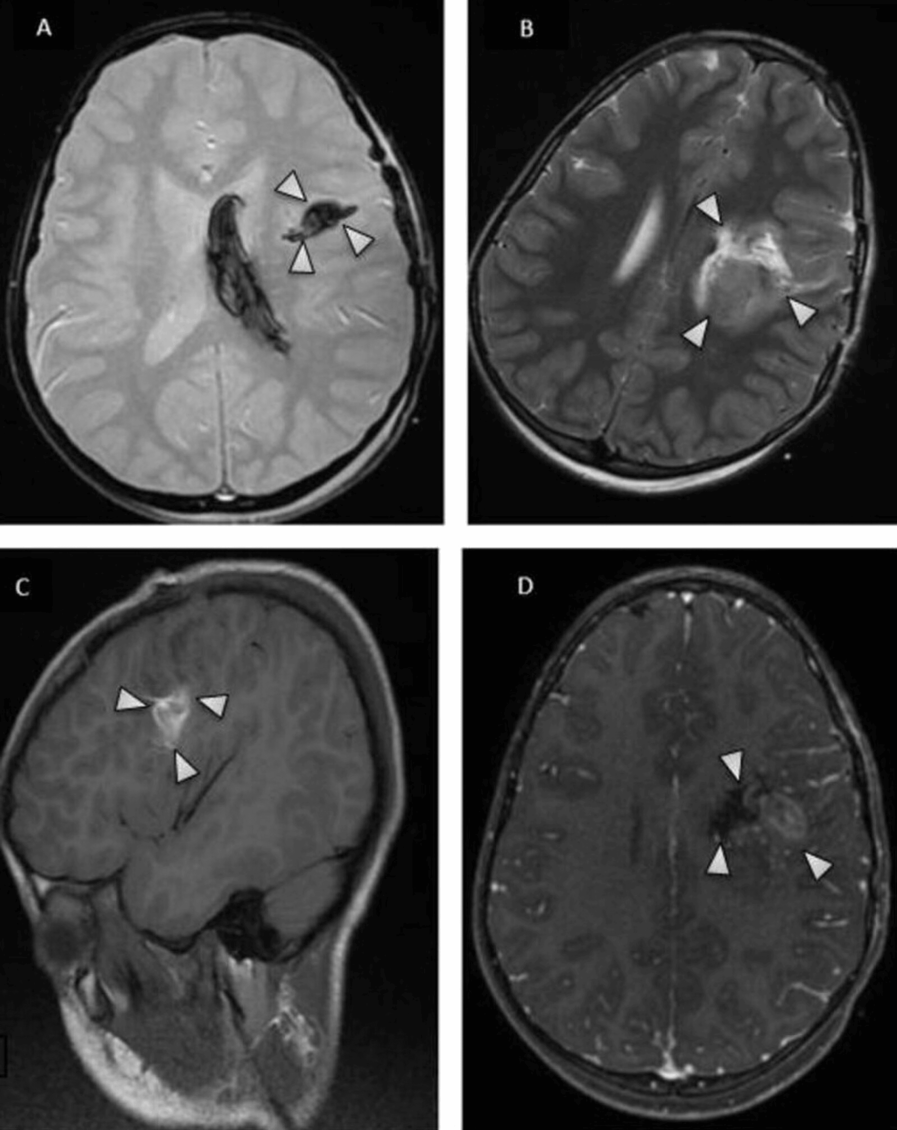
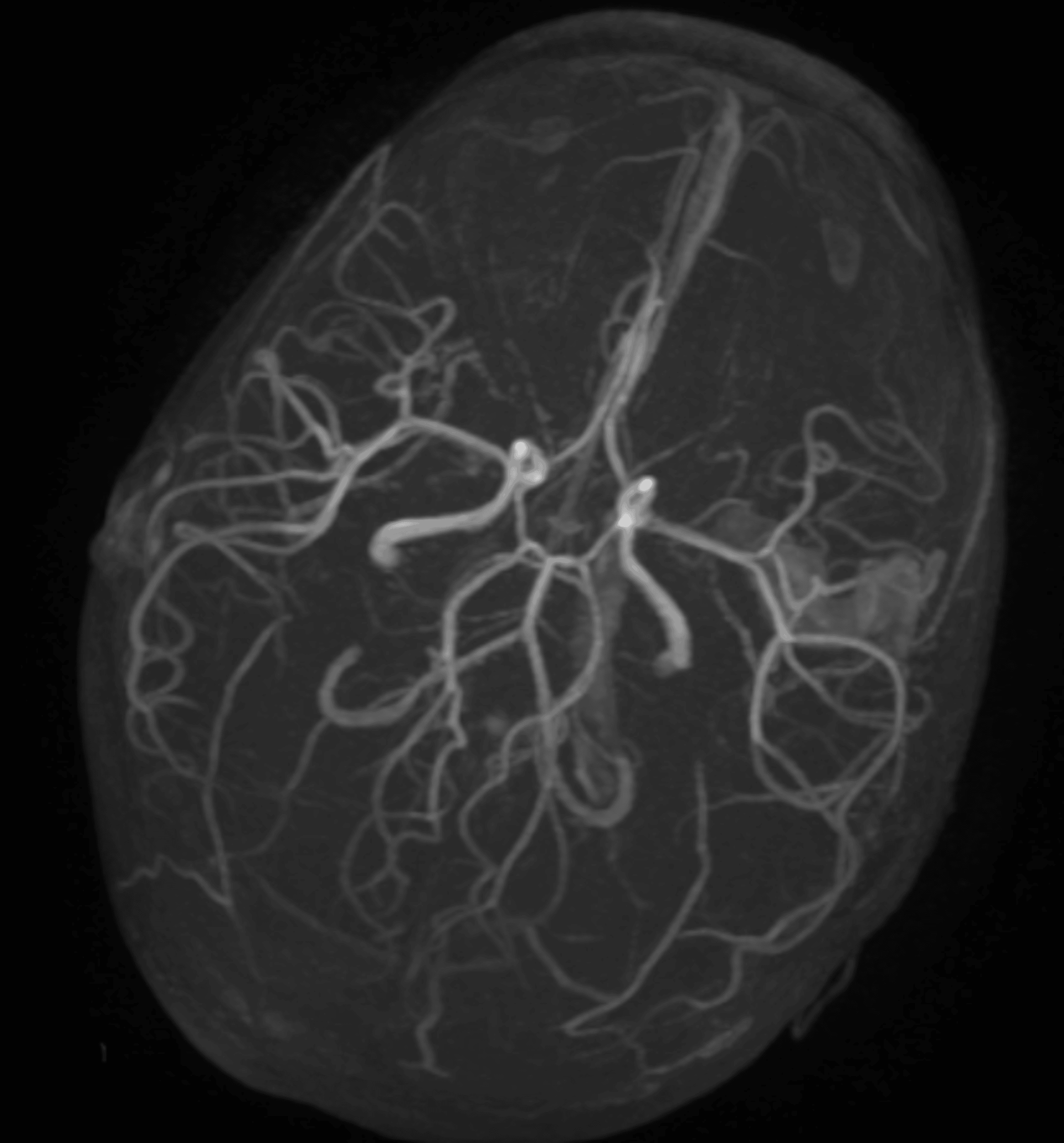
The postoperative course in the intensive care unit was favorable. Brain magnetic resonance imaging (MRI) performed on postoperative day three demonstrated an evolving intraparenchymal hematoma with susceptibility-related signal loss on the T2*-weighted sequence and heterogeneous signal characteristics on T1- and T2-weighted sequences, without appreciable post-contrast enhancement (Figure 1). Magnetic resonance angiography (MRA) was also obtained as part of the vascular assessment, using a three-dimensional time-of-flight (3D TOF) sequence. It demonstrated preserved patency of the major intracranial arteries of the circle of Willis, without an apparent intracranial aneurysm or significant arterial stenosis (Figure 2). This examination did not establish the underlying cause of the hemorrhage. Arterial 3D TOF MRA does not exclude cerebral venous thrombosis or all small or occult vascular lesions. Dedicated MR venography or CT venography and catheter-based digital subtraction angiography (DSA) were not documented in the available records. Sedation was discontinued at hour 48, with recovery of consciousness and extubation on postoperative day three. After sedation withdrawal and extubation, right hemiplegia and expressive aphasia were documented. The available records do not establish the precise onset of these focal deficits relative to the initial presentation and surgery. She was transferred to the neurosurgery department on the fourth postoperative day for continued specialized care and initiation of multidisciplinary rehabilitation.
Functional follow-up was conducted jointly by physiotherapy, occupational therapy, speech-language therapy, and psychomotor therapy, with formal assessments at one, three, and six months. At one month, the child was conscious, alert, and actively participating in rehabilitation sessions. Right hemiparesis (recovering from the initial hemiplegia) predominated in the upper limb, with marked distal weakness limiting fine motor control. The right lower limb had recovered more rapidly, allowing standing with assistance and a few assisted steps. Language impairment was predominantly expressive, characterized by reduced verbal fluency and word-finding difficulty, while comprehension of simple instructions was broadly preserved. Physiotherapy focused on postural control, transfers, standing, and gait retraining. Occupational therapy targeted functional use of the right upper limb. Speech-language therapy addressed comprehension, verbal expression, and naming. Psychomotor therapy focused on body schema, coordination, and motor planning.
At three months, motor recovery had progressed substantially. Transfers were independent, and the child was able to walk on level ground, initially under supervision and subsequently without assistance over short distances, with a discreet residual limp. In the right upper limb, proximal motor control had improved and voluntary grasp was possible, although persistent distal weakness limited fine motor activities. Gross grasp had returned sufficiently to permit functional object handling, but the available records did not quantify grip strength, pinch strength, or standardized dexterity performance. Language had also improved considerably, allowing functional oral communication. However, word-finding difficulty and slowed speech production persisted during more complex verbal tasks. Rehabilitation therefore progressed toward dynamic balance and selective coordination of the right limbs, activities of daily living and school-related tasks, language fluency and syntax, and visuomotor coordination and praxis.
At six months, overall functional recovery was favorable. The child was independent in the main activities of daily living and walked independently without an assistive device. Gait remained mildly asymmetric, with some fatigability during sustained activity. The right upper limb was used for most bimanual activities, although reduced dexterity persisted during fine graphic tasks. Spontaneous communication was functional in everyday situations, with residual difficulty in lexical access and complex verbal formulation. Continued speech-language therapy was recommended in preparation for progressive return to school. No validated standardized pediatric functional outcome measure was documented in the available follow-up records; the functional trajectory is therefore reported descriptively. The functional trajectory at one, three, and six months is summarized in Table 2.
The functional trajectory therefore demonstrated substantial recovery while leaving mild residual motor and language deficits at six months. Continued multidisciplinary follow-up was maintained because neurological, cognitive, language, and school-related difficulties may persist or become more apparent during recovery from pediatric intracerebral hemorrhage.
Discussion
Pediatric intracerebral hemorrhage is an uncommon but potentially severe neurological emergency. Its clinical presentation may be nonspecific, with seizures, vomiting, headache, and impaired consciousness potentially mimicking infectious or metabolic conditions [2,3]. In the present case, a brief episode of hyperthermia preceded the neurological deterioration, but fever was not persistent during transfer or admission. The subsequent identification of an intracerebral hematoma emphasizes the importance of early neuroimaging in children with acute unexplained neurological impairment. Although fever can occur in infectious neurological disorders, its presence at symptom onset should not prevent consideration of intracranial hemorrhage when seizures and altered consciousness are present. In this patient, the absence of persistent fever and the subsequent imaging findings supported a structural neurological cause. Nevertheless, the observation from a single case cannot establish a causal relationship between transient fever and hemorrhage.
The etiological spectrum of pediatric spontaneous intracranial hemorrhage differs from that of adult intracerebral hemorrhage. A systematic review including 7,931 children and adolescents found that vascular etiologies represented 43.08% of identified etiologies and risk factors, with arteriovenous malformations accounting for 70.99% of vascular causes [2]. Consequently, vascular imaging has an important role in the evaluation of pediatric intracerebral hemorrhage. Current international consensus recommendations recognize MRA and computed tomography angiography as appropriate noninvasive techniques for the initial vascular assessment, while digital subtraction angiography may be required when noninvasive imaging is inconclusive or when an occult vascular lesion remains suspected [4]. In our patient, postoperative MRI was performed on postoperative day three. The examination demonstrated the expected evolving hematoma without appreciable post-contrast enhancement. Vascular imaging using 3D TOF MRA demonstrated patent major intracranial arteries without an apparent aneurysm or significant stenosis.
The 3D TOF technique is a noninvasive MRA method that provides flow-related visualization of intracranial arteries. A meta-analysis evaluating 3D TOF MRA for intracranial aneurysm detection supports its role in noninvasive arterial assessment but does not establish its ability to exclude arteriovenous malformations or other occult vascular shunts [5]. The negative arterial 3D TOF MRA must therefore be interpreted within the limits of the technique. It did not establish a definitive cause and cannot exclude cerebral venous thrombosis, a small arteriovenous malformation, or another occult vascular lesion. Dedicated MR venography or CT venography was not documented in the available records. In addition, catheter-based DSA was not performed during the reported follow-up period. The absence of an abnormality on the available MRA should therefore be described as the absence of an apparent arterial lesion rather than definitive exclusion of a vascular cause.
The International Pediatric Stroke Organization consensus statement on acute evaluation and management recommends DSA in children whose initial vascular work-up does not reveal an obvious cause, unless a specific diagnosis makes angiography unnecessary [4]. Repeat vascular assessment may also be required when an initial investigation is negative and suspicion persists. No DSA or delayed vascular imaging was documented during the six-month follow-up in this case. This represents a substantial limitation of the etiological evaluation. A pediatric neurovascular team should consider further investigation, including arterial and venous imaging and delayed reassessment after hematoma resolution, according to the clinical context and local resources. Because the cause remains undetermined, the possibility of an occult lesion and the associated risk of recurrence cannot be resolved from the available data.
The acute management of pediatric intracerebral hemorrhage requires rapid stabilization, maintenance of adequate cerebral perfusion, control of ventilation and oxygenation, management of intracranial pressure when available, and early neurosurgical assessment [4]. In the present case, the child was intubated before transfer because of severe impairment of consciousness. Mechanical ventilation, head elevation, invasive blood pressure monitoring, and osmotherapy were used during the acute phase. Intracranial pressure monitoring was not available in our institution. The absence of direct intracranial pressure monitoring represents an important limitation, although clinical and radiological surveillance allowed subsequent sedation withdrawal and neurological assessment. Emergency surgical evacuation was undertaken in the context of severe neurological impairment, intraventricular extension, and reported mass effect associated with the intracerebral hemorrhage. Current pediatric consensus recommendations emphasize individualized neurosurgical decision-making according to hematoma location, mass effect, neurological status, and the suspected underlying etiology [4]. In the emergency setting, treatment may necessarily precede complete etiological characterization, particularly when mass effect creates an immediate threat to neurological function.
The original CT images and quantitative measurements were unavailable, and the exact anatomical epicenter and hematoma volume could not be independently confirmed. The operative report was also insufficiently detailed in the available documentation to establish the precise surgical approach, intraoperative findings concerning a possible vascular lesion, external ventricular drain placement, or histopathological results. These limitations restrict the extent to which the indication and technical details of surgery can be discussed retrospectively.
Seizures are also clinically important in pediatric intracerebral hemorrhage. A recent retrospective study of 148 children with spontaneous intracranial hemorrhage found that seizures at onset were independently associated with poor three-month neurological outcome, although the authors emphasized the exploratory nature of these findings and the need for external validation [6]. In the present case, seizures were the initial manifestation, but no subsequent epileptic disorder was documented during the six-month follow-up. The available records did not provide sufficient detail regarding acute antiseizure medication, electroencephalographic monitoring, or the total seizure burden; therefore, no conclusion can be drawn about the effect of a specific seizure-management strategy. Continued neurological surveillance remains appropriate.
Functional outcome after pediatric intracerebral hemorrhage is heterogeneous. In a cohort and meta-analysis of pediatric intracerebral hemorrhage, 57.4% of children had a favorable clinical outcome at follow-up, while pooled complete recovery was 27% and the aggregate case-fatality rate was 17.3% [7]. These findings emphasize that survival does not necessarily imply complete neurological recovery. Our patient’s functional trajectory illustrates this distinction. Recovery was progressive rather than immediate, with earlier improvement in lower-limb function followed by slower recovery of upper-limb dexterity and language. At six months, she was independent in the principal activities of daily living and walked without an assistive device, but mild right-sided motor impairment and language difficulties persisted. Contemporary consensus recommendations emphasize the importance of multidisciplinary rehabilitation and long-term follow-up after pediatric intracerebral hemorrhage, including attention to motor, communication, cognitive, behavioral, educational, and psychosocial outcomes [8]. The rehabilitation program in our patient therefore involved physiotherapy, occupational therapy, speech-language therapy, and psychomotor therapy, with progressive adaptation of goals according to functional recovery.
The rehabilitation findings were documented descriptively. Standardized measures such as pediatric activities-of-daily-living scales, manual ability classifications, and validated language or cognitive assessments were not recorded in the available follow-up data. Consequently, the magnitude of recovery cannot be quantified using standardized scores, and the reported functional trajectory should be interpreted as a clinical description rather than a validated outcome measurement.
This report has several limitations. First, the original CT images were obtained at the referring facility and were not available for inclusion because the source image files could not be retrieved from the patient’s available medical records. The CT findings reported in this manuscript were based on the clinical documentation accompanying the patient. The exact hemorrhage location, dimensions, volume, degree of midline shift, and presence or absence of hydrocephalus could not be independently verified. Second, intracranial pressure monitoring was not available during the acute phase. Third, the initial neurological examination was potentially confounded by sedative and neuromuscular-blocking medication administered before transfer. Fourth, the etiological work-up was incomplete: no dedicated venous imaging, DSA, or delayed vascular follow-up was documented during the six-month observation period. An occult vascular lesion or cerebral venous thrombosis therefore cannot be excluded. Finally, the six-month functional trajectory represents a single-patient observation without prospectively collected standardized pediatric outcome scores and should not be generalized to the broader pediatric intracerebral hemorrhage population.
Conclusions
Pediatric intracerebral hemorrhage should be considered in children presenting with acute impaired consciousness and seizures, even when fever is transient or absent. Rapid stabilization, early neuroimaging, multidisciplinary neurosurgical and intensive care management, and a comprehensive etiological evaluation with appropriate vascular follow-up are important components of care.
In this case, the etiology remains undetermined. The absence of an apparent lesion on 3D TOF MRA should not be interpreted as definitive exclusion of an underlying vascular cause, particularly because dedicated venous imaging, catheter-based DSA, and delayed vascular reassessment were not documented. Further etiological investigation should be considered according to specialist assessment and available resources.
References
- Haizel-Cobbina J, Koester SW, Arafeh Y, et al.: Global pediatric stroke epidemiology: a systematic review of the literature and meta-analysis. J Child Neurol. 2026, 41:1220-8. 10.1177/08830738261441084
- Ciochon UM, Bindslev JB, Hoei-Hansen CE, Truelsen TC, Larsen VA, Nielsen MB, Hansen AE: Causes and risk factors of pediatric spontaneous intracranial hemorrhage: a systematic review. Diagnostics (Basel). 2022, 12:10.3390/diagnostics12061459
- Sporns PB, Psychogios MN, Fullerton HJ, Lee S, Naggara O, Boulouis G: Neuroimaging of pediatric intracerebral hemorrhage. J Clin Med. 2020, 9:10.3390/jcm9051518
- Boulouis G, Fox CK, Waak M, et al.: Pediatric intracerebral hemorrhage management-consensus statement of the International Pediatric Stroke Organization-part 1: acute phase and workup. J Am Heart Assoc. 2026, 15:e039594. 10.1161/JAHA.124.039594
- HaiFeng L, YongSheng X, YangQin X, Yu D, ShuaiWen W, XingRu L, JunQiang L: Diagnostic value of 3D time-of-flight magnetic resonance angiography for detecting intracranial aneurysm: a meta-analysis. Neuroradiology. 2017, 59:1083-92. 10.1007/s00234-017-1905-0
- Hou W, Wang S, Fang J: Etiology and early clinical predictors of neurological outcomes in pediatric spontaneous intracranial hemorrhage: a retrospective exploratory study. Front Neurol. 2025, 16:1750475. 10.3389/fneur.2025.1750475
- Boulouis G, Stricker S, Benichi S, et al.: Mortality and functional outcome after pediatric intracerebral hemorrhage: cohort study and meta-analysis. J Neurosurg Pediatr. 2021, 27:661-7. 10.3171/2020.9.PEDS20608
- Mrakotsky C, Mailo JA, Chevignard M, et al.: Pediatric intracerebral hemorrhage management-consensus statement of the International Pediatric Stroke Organization-part 2: outcomes, rehabilitation, and transition to adulthood. J Am Heart Assoc. 2026, 15:e039595.