

Authors: Mavridou P, Kitsakou P, Exarchos C, et al.
Cureus 18(7): e112936. doi:10.7759/cureus.112936
Abstract
Negative pressure pulmonary edema (NPPE) is a rare, life-threatening, yet rapidly reversible complication of acute upper airway obstruction. We present the case of a 17-year-old healthy male who underwent an emergency appendectomy under general anesthesia. Immediately following extubation, he developed severe respiratory distress and desaturation (SpO₂ 70%) due to presumed glossoptosis and reactive laryngospasm. Despite initial stabilization, he deteriorated in the Post-anesthesia Care Unit, exhibiting hypoxemia and pink frothy sputum. Arterial blood gases confirmed acute respiratory acidosis, while chest radiography revealed diffuse bilateral alveolar opacities. A completely normal point-of-care ultrasound (POCUS) served as a valuable adjunct to effectively rule out cardiogenic edema. The patient was managed conservatively with supplemental oxygen and nebulized bronchodilators, while loop diuretics were deliberately avoided. He showed rapid clinical improvement, weaning to room air within six hours, with complete radiological resolution by the next morning. While NPPE is a recognized complication, the educational novelty of this case lies in highlighting the efficacy of targeted conservative management. It underscores the adjunctive role of POCUS in safely avoiding inappropriate diuresis and emphasizes the paramount need for continuous vigilance following extubation.
Introduction
Negative pressure pulmonary edema (NPPE), also known as post-obstructive pulmonary edema, is a rare but potentially life-threatening complication of acute upper airway obstruction. It is well-documented yet frequently underdiagnosed in clinical practice [1]. The condition typically occurs when a patient makes a forceful, involuntary inspiratory effort against a closed glottis (vocal cords) or an obstructed airway (the Müller maneuver). This physiological mechanism drastically increases negative intrapleural pressure (the pressure within the chest cavity), which, in turn, augments venous return, increases pulmonary capillary hydrostatic pressure, and ultimately leads to rapid transudation (leakage) of fluid into the alveolar space (the air sacs of the lungs) [2,3].
NPPE occurs in approximately 0.05% to 0.1% of all procedures performed under general anesthesia [4]. It most commonly develops secondary to acute upper airway obstruction, such as post-extubation laryngospasm (which has an overall incidence of approximately 1% in general anesthesia) or posterior displacement of the tongue (glossoptosis) due to a reduced level of consciousness. This particularly affects young, muscular, and otherwise healthy individuals capable of generating high intrathoracic pressure [5]. While the clinical presentation is dramatic-often characterized by acute hypoxemia, respiratory distress, and pink frothy sputum-the condition is highly reversible if recognized and managed promptly. The primary diagnostic challenge lies in differentiating NPPE from cardiogenic pulmonary edema, aspiration pneumonitis, and anaphylaxis in the immediate post-extubation period [6]. In this context, bedside ultrasound has emerged as a crucial adjunctive tool to rapidly narrow the differential diagnosis. We present a case of NPPE in a 17-year-old male following an emergency appendectomy, emphasizing the typical clinical course, the necessity of targeted differential diagnosis, and the paramount importance of continuous vigilance even after initial post-extubation stabilization.
Case Presentation
Patient presentation and surgical course
A 17-year-old male patient, weighing 71 kg with a height of 175 cm (body mass index: 23.2 kg/m²), with no significant past medical history or known allergies (American Society of Anesthesiologists physical status I), presented to the emergency department reporting worsening periumbilical pain that migrated to the right lower quadrant, accompanied by anorexia. According to the official report provided by the radiologists, the preoperative abdominal ultrasound revealed a dilated (up to 1 cm), thick-walled appendix with periappendiceal fluid and local fat stranding, confirming acute appendicitis. Initial laboratory investigations performed in the emergency department during acute triage showed leukocytosis (15.62 k/μL) and a normal baseline high-sensitivity troponin I (5.4 ng/L) (Table 1).
Based on the combined clinical, ultrasonographic, and laboratory findings, the patient was scheduled for an emergency open appendectomy under general anesthesia. Routine monitoring (electrocardiography, non-invasive blood pressure, pulse oximetry, and Train-of-Four (TOF) monitoring) was applied. Pharmacological prophylaxis for postoperative nausea and vomiting (PONV) and aspiration was administered, including intravenous dexamethasone 8 mg, ondansetron, and omeprazole. General anesthesia was induced using intravenous propofol 200 mg, rocuronium 70 mg, and fentanyl 200 µg. The trachea was intubated without difficulty using a 7.0 mm endotracheal tube, and anesthesia was maintained with sevoflurane in an oxygen/air mixture. The open appendectomy lasted 55 minutes and was completed without intraoperative hemodynamic or respiratory complications.
Immediate post-extubation crisis
During the emergence phase, neuromuscular blockade was fully reversed with intravenous sugammadex 150 mg (approximately 2 mg/kg), and full reversal was objectively documented by a TOF ratio greater than 0.9. The patient’s oropharynx was suctioned, and an awake extubation was performed. Immediately following extubation, the patient exhibited signs of acute upper airway obstruction, presumably due to posterior displacement of the tongue associated with reduced wakefulness, rapidly followed by reactive laryngospasm. He rapidly developed acute respiratory distress, audible bronchospasm, and profound oxygen desaturation, with peripheral capillary oxygen saturation (SpO₂) dropping to 70%. Immediate bag-valve-mask positive pressure ventilation with 100% oxygen was initiated. Concurrently, intravenous hydrocortisone (500 mg) was administered specifically to mitigate the severe reactive bronchospasm and airway inflammation. These initial rescue measures successfully broke the laryngospasm and temporarily restored oxygenation, elevating the SpO₂ to 98%.
Secondary deterioration and diagnostic workup
The patient was subsequently transferred to the Post-anesthesia Care Unit (PACU) for continuous monitoring. Shortly after arrival in the PACU, he experienced a secondary clinical deterioration. He became tachypneic, distressed, and began coughing, producing characteristic pink, frothy sputum. His SpO₂ declined again to 87% on room air. An arterial blood gas (ABG) analysis performed on room air revealed acute respiratory acidosis and hypoxemia: pH 7.33, PaCO₂ 50 mmHg, PaO₂ 65 mmHg, HCO₃⁻ 26 mEq/L, and SaO₂ 91%.
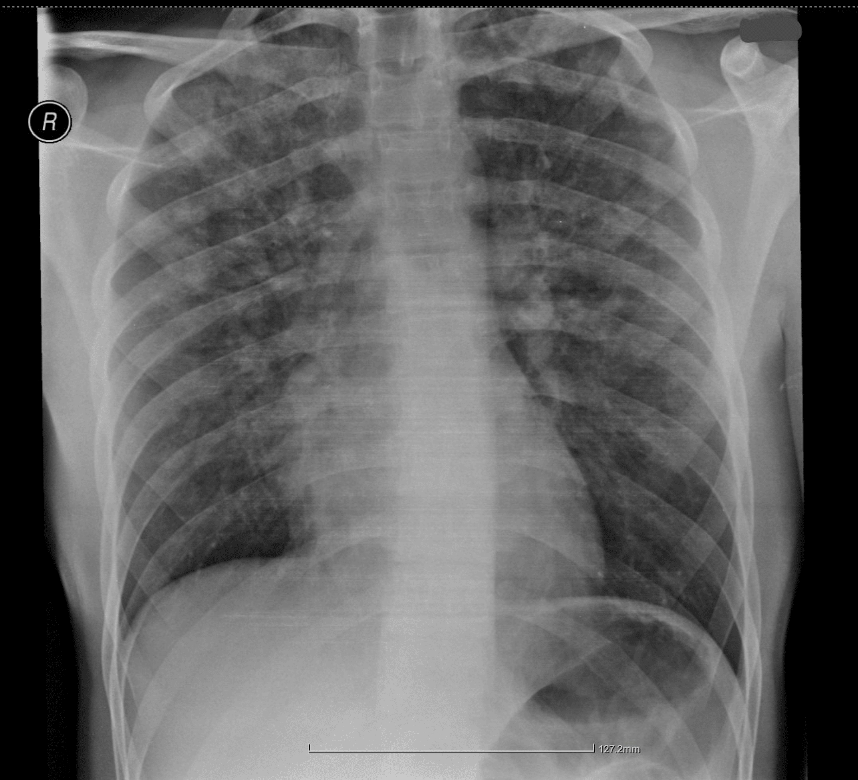
Based on the clinical presentation, NPPE was strongly suspected. Treatment with supplemental oxygen via a face mask and nebulized bronchodilators was initiated immediately. A pulmonology consultation was requested; the consulting pulmonologist noted bilateral wheezing, prolonged expiration, and basal crackles. Following the administration of nebulized ipratropium bromide/fenoterol, nebulized budesonide, and an additional dose of intravenous methylprednisolone (40 mg), the auscultatory findings improved, and a chest X-ray was recommended. A portable chest radiograph demonstrated diffuse, bilateral, ill-defined opacities primarily in the central and lower zones, lacking pleural effusions, and featuring a normal cardiac silhouette (Figure 1).
To definitively rule out cardiogenic pulmonary edema, a cardiology consultation was requested. Cardiac point-of-care ultrasound (POCUS) demonstrated preserved biventricular systolic function, no significant valvular abnormalities, and a normal-sized inferior vena cava (IVC) with preserved respiratory variation. These findings did not support elevated right-sided filling pressures or an overt cardiogenic cause of respiratory deterioration. Furthermore, a post-crisis high-sensitivity troponin I level was evaluated at 6.7 ng/L (reference range 0 – 34.2 ng/L), showing no significant elevation from the preoperative baseline, while total creatine kinase (CK) showed a mild postoperative elevation to 272 IU/L. The combination of a normal POCUS and negative serial troponins strongly supported the diagnosis of non-cardiogenic NPPE.
Targeted management and resolution
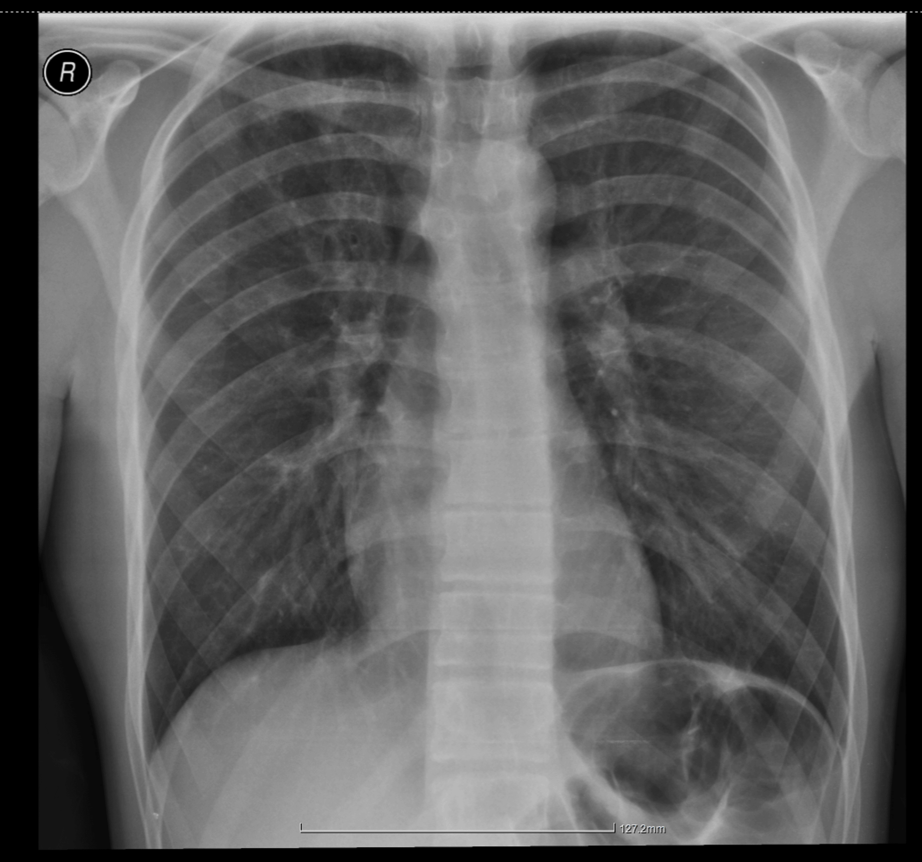
The patient was transferred to the surgical ward on continuous face-mask oxygen therapy; attempts to remove the mask initially resulted in rapid desaturation. After six hours of conservative management, his respiratory status significantly improved. He became completely asymptomatic, the coughing ceased, and he maintained an SpO₂ of 97% on room air. He remained clinically stable overnight, and his condition was deemed fully resolved. A follow-up chest radiograph performed on postoperative day 1 (POD 1) (approximately 20 hours after the onset of the acute respiratory crisis) demonstrated complete resolution of the pulmonary opacities (Figure 2).
The patient continued to recover uneventfully on the ward and was safely discharged home on the third postoperative day in excellent clinical condition.
Discussion
Pathophysiology and classification
NPPE is traditionally classified into two distinct clinical entities: Type I and Type II (Table 2) [7-9]. The present case represents a classic presentation of Type I NPPE, developing acutely after extubation in a healthy adolescent.
The pathophysiology of Type I NPPE is driven by the Müller maneuver, a vigorous inspiratory effort against a closed glottis. In young, healthy individuals with significant diaphragmatic strength, this effort can generate negative intrathoracic pressures reaching -100 cm H₂O, compared to a normal resting pressure of -4 to -9 cm H₂O [9]. This extreme negative intrathoracic pressure significantly increases venous return to the right atrium while simultaneously increasing left ventricular afterload. The resulting hydrostatic gradient forces rapid transudation of fluid from the pulmonary capillaries into the interstitium and alveolar spaces, presenting clinically with hypoxia and the hallmark pink, frothy sputum [2,10].
Diagnostic approach and the role of POCUS
In the immediate postoperative setting, diagnosing NPPE requires distinguishing its unique hydrostatic mechanism (driven by extreme negative intrathoracic pressure) from other causes of acute hypoxemia. These include the inflammatory capillary leak characteristic of aspiration pneumonitis (Mendelson’s syndrome) or anaphylaxis, and the primary pump failure or volume overload seen in cardiogenic pulmonary edema [3,11]. Aspiration was deemed highly unlikely in our patient as there was no evidence of gastric regurgitation during extubation, and the rapid clinical improvement over a few hours is inconsistent with the typical prolonged course of aspiration pneumonitis. Furthermore, the completely normal bedside echocardiogram provided by the consulting cardiologist was pivotal in confirming the diagnosis. Serial negative high-sensitivity troponin levels conclusively ruled out myocardial ischemia or stress-induced cardiomyopathy, while the mild postoperative elevation in total creatine kinase (CK) was an expected physiological response attributed to the surgical tissue trauma and the intense work of breathing during the acute airway obstruction. POCUS has emerged as an invaluable tool in these acute settings; it quickly rules out primary cardiac dysfunction or gross volume overload, obviating the need for delayed laboratory biomarkers such as proBNP, and preventing the erroneous and potentially harmful administration of loop diuretics, which could exacerbate hypovolemia in a recently operated patient [12].
Targeted conservative management
The management of NPPE is primarily supportive and aims to correct hypoxemia while the fluid is cleared by the pulmonary lymphatic system. Ensuring airway patency and providing supplemental oxygen or non-invasive positive pressure ventilation are usually sufficient [4,7]. A recent case report published in Cureus by Jagadish et al. similarly highlighted the efficacy of conservative management in a young patient developing NPPE following an appendectomy [13]. However, while their successful treatment regimen included the administration of intravenous furosemide, our decision to strictly avoid loop diuretics was a deliberate, evidence-based choice. Unless fluid overload is definitively proven, diuresis is generally contraindicated as it can exacerbate postoperative hypovolemia and lead to hemodynamic instability. The integration of POCUS in our case provided the clinical confidence to safely withhold diuretics. Furthermore, while systemic corticosteroids are not indicated for the treatment of hydrostatic pulmonary edema itself, the intravenous hydrocortisone in our patient was administered specifically to mitigate the severe reactive bronchospasm and localized upper airway edema secondary to the violent laryngospasm. Additionally, the use of nebulized β2-agonists (fenoterol/ipratropium) served a dual purpose: it successfully relieved the auscultated bronchospasm and, as recent evidence suggests, β2-adrenergic stimulation actively accelerates alveolar fluid clearance by upregulating transepithelial sodium transport mechanisms [14].
The need for continuous vigilance
Crucially, this case underscores the necessity of continuous vigilance during the entire perioperative period. While extubation is often viewed as the conclusion of airway management, the immediate post-extubation phase and the subsequent hours in the PACU remain high-risk periods for the development of delayed respiratory complications [1,5]. Furthermore, since residual neuromuscular blockade is a well-known precipitant of upper airway obstruction, considering the re-assessment of relaxant reversal, even after initial objective documentation of recovery in the operating room, adds significant value during the immediate postoperative period to prevent such catastrophic events. As demonstrated in our patient, even after an initial recovery from laryngospasm, NPPE can manifest subsequently, necessitating careful and continuous monitoring. Consistent with the literature, conservative management facilitated the rapid clearance of pulmonary infiltrates and a complete clinical recovery within 24 hours [6,11].
Conclusions
NPPE is a severe but highly reversible complication of acute upper airway obstruction, most frequently triggered by laryngospasm during or immediately after extubation. Prevention primarily relies on meticulous airway management, ensuring complete reversal of neuromuscular blockade, and performing extubation only when the patient is fully awake and able to protect their airway. Anesthesiologists and PACU staff must maintain a high index of suspicion and practice continuous vigilance, even after initial airway stabilization, especially when dealing with young, muscular patients capable of generating immense negative intrathoracic pressure. Rapid differentiation from cardiogenic pulmonary edema is essential, with bedside echocardiography (POCUS) serving as a highly valuable adjunct to clinical assessment. Prompt recognition and supportive management, emphasizing oxygenation and positive airway pressure rather than aggressive diuresis, ensure full clinical and radiological recovery, typically within 24 hours.
References
- Fremont RD, Kallet RH, Matthay MA, Ware LB: Postobstructive pulmonary edema: a case for hydrostatic mechanisms. Chest. 2007, 131:1742-6. 10.1378/chest.06-2934
- Wu J, Yuan H, Guo Z, Feng Q, Ma J: Negative pressure pulmonary edema: a case report and literature review. Front Med (Lausanne). 2024, 11:1467541. 10.3389/fmed.2024.1467541
- Bhattacharya M, Kallet RH, Ware LB, Matthay MA: Negative-pressure pulmonary edema. Chest. 2016, 150:927-33. 10.1016/j.chest.2016.03.043
- Luo M, Li M, Qin Z: Negative pressure pulmonary edema resulting from upper airway obstruction during the post-anesthesia recovery period: a case series and literature review. BMC Anesthesiol. 2025, 25:125. 10.1186/s12871-025-02998-z
- Guru PK, Agarwal A, Pimentel M, McLaughlin DC, Bansal V: Postoperative pulmonary edema conundrum: a case of negative pressure pulmonary edema. Case Rep Crit Care. 2018, 2018:1584134. 10.1155/2018/1584134
- Ma J, Liu T, Wang Q, et al.: Negative pressure pulmonary edema (review). Exp Ther Med. 2023, 26:455. 10.3892/etm.2023.12154
- Lemyze M, Mallat J: Understanding negative pressure pulmonary edema. Intensive Care Med. 2014, 40:1140-3. 10.1007/s00134-014-3307-7
- Park H, Nam S, Jang YJ, Ku S, Choi SS: Negative pressure pulmonary edema in a patient undergoing open rhinoplasty: a case report. Medicine (Baltimore). 2021, 100:e24240. 10.1097/MD.0000000000024240
- Udeshi A, Cantie SM, Pierre E: Postobstructive pulmonary edema. J Crit Care. 2010, 25:508.e1-5. 10.1016/j.jcrc.2009.12.014
- Costa D, Teles L, Prucha Leite N, Tadeu J: When the unexpected happens: diffuse alveolar hemorrhage in negative-pressure pulmonary edema. Cureus. 2025, 17:e87323. 10.7759/cureus.87323
- Bhaskar B, Fraser JF: Negative pressure pulmonary edema revisited: pathophysiology and review of management. Saudi J Anaesth. 2011, 5:308-13. 10.4103/1658-354X.84108
- Silva A, Furtado I, Grenho B, Isidoro M: Point of care ultrasound-diagnostic approach of an atypical negative pressure pulmonary oedema. Turk J Anaesthesiol Reanim. 2022, 50:306-8. 10.5152/TJAR.2022.21365
- Jagadish A, Ross JM, Gibson B: Conservative management of negative pressure pulmonary edema: a case report. Cureus. 2026, 18:e103213. 10.7759/cureus.103213
- Matthay MA: Resolution of pulmonary edema. Thirty years of progress. Am J Respir Crit Care Med. 2014, 189:1301-8. 10.1164/rccm.201403-0535OE