

Authors: Khan A et al
Cureus 18(10): e118609. doi:10.7759/cureus.118609
Abstract
A 34-year-old man was admitted with fever, cough, chest pain, headache, and acute confusion, raising an initial suspicion of meningoencephalitis. However, two sets of blood cultures obtained before empirical antibiotic therapy grew methicillin-sensitive Staphylococcus aureus (MSSA), and an electrocardiogram showed complete heart block. His condition deteriorated rapidly, progressing to respiratory failure and cardiogenic shock. Cardiac imaging demonstrated multivalvular infective endocarditis with severe mitral regurgitation secondary to perforation of the mitral leaflet. Emergency valve surgery was undertaken, followed by permanent pacing. This case emphasises the importance of early cardiac assessment in patients with systemic infection and conduction abnormalities, as it highlights an atypical presentation of infective endocarditis mimicking neurological infection.
Introduction
Infective endocarditis (IE) is a potentially life-threatening infection of the endocardial surface, most commonly involving the cardiac valves, with an estimated annual incidence of 3-10 cases per 100,000 population. It is predominantly caused by Gram-positive organisms, including Staphylococcus aureus, Streptococci, and Enterococci. S. aureus is particularly associated with aggressive valvular destruction and systemic complications. Treatment involves prolonged pathogen-directed antimicrobial therapy, with surgery required in patients with severe valvular dysfunction or uncontrolled infection [1,2].
The clinical presentation of IE is variable, ranging from fever and malaise to heart failure, systemic embolisation, and septic shock. Neurological manifestations, including stroke, encephalopathy, and cerebral abscesses, may be the initial presentation, occasionally mimicking meningoencephalitis and delaying diagnosis [1,2].
Conduction abnormalities are less common but clinically important. New atrioventricular block, particularly complete heart block, may indicate peri-annular extension of infection involving the cardiac conduction system and should prompt urgent echocardiographic assessment. Multivalvular IE is also uncommon and is associated with greater diagnostic and therapeutic complexity, often requiring surgical intervention [1,3].
We report a 34-year-old man who presented with fever, headache, and confusion, initially raising suspicion of meningoencephalitis. Subsequent investigations revealed methicillin-sensitive Staphylococcus aureus (MSSA) bacteraemia, persistent complete heart block, and IE involving the mitral, tricuspid, and pulmonary valves. The clinical course was complicated by mitral leaflet perforation, severe mitral regurgitation, and cardiogenic shock, requiring emergency surgery and permanent pacemaker implantation. This case highlights the importance of recognising atypical presentations of IE and investigating unexplained conduction abnormalities in patients with systemic infection.
Case Presentation
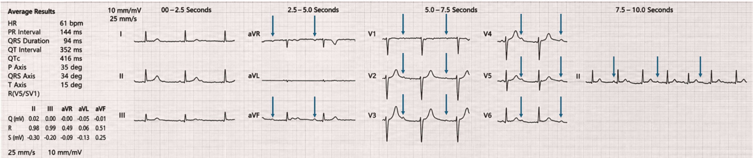
A man in his 30s presented to the Emergency Department with a one-week history of fever and cough, followed by three days of chest pain, headache, and confusion. On admission, he was febrile (39.4°C), bradycardic (43 beats/min), and tachypnoeic (26 breaths/min), with an oxygen saturation of 85% on room air, a blood pressure of 113/76 mmHg, and a Glasgow Coma Scale score of 14/15 (Table 1). Initial investigations demonstrated markedly elevated inflammatory markers, including a white cell count of 17.3 × 10⁹/L and C-reactive protein of 384 mg/L, alongside an elevated troponin I of 1,112 ng/L (Table 2). Electrocardiography demonstrated complete heart block (Figure 1).
The initial differential diagnosis included meningoencephalitis. Further history, including collateral information from relatives, revealed recent wrist injuries sustained while constructing a shed door for pets, although there was no reported exposure to animal secretions. Lumbar puncture demonstrated cerebrospinal fluid pleocytosis (35 × 10⁶ cells/L), with 60% lymphocytes and 40% neutrophils. Cerebrospinal fluid culture subsequently showed no growth. These findings initially supported the possibility of meningoencephalitis.
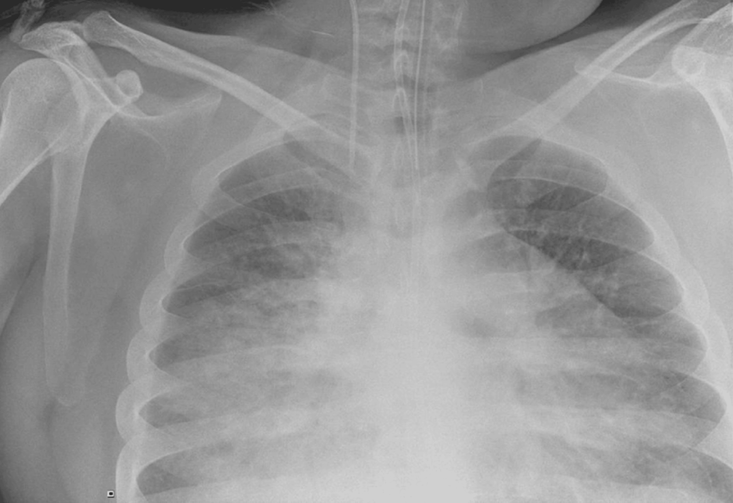
During admission, the patient’s respiratory condition deteriorated, with increasing oxygen requirements. Chest radiography demonstrated bilateral interstitial changes suggestive of pulmonary oedema (Figure 2).
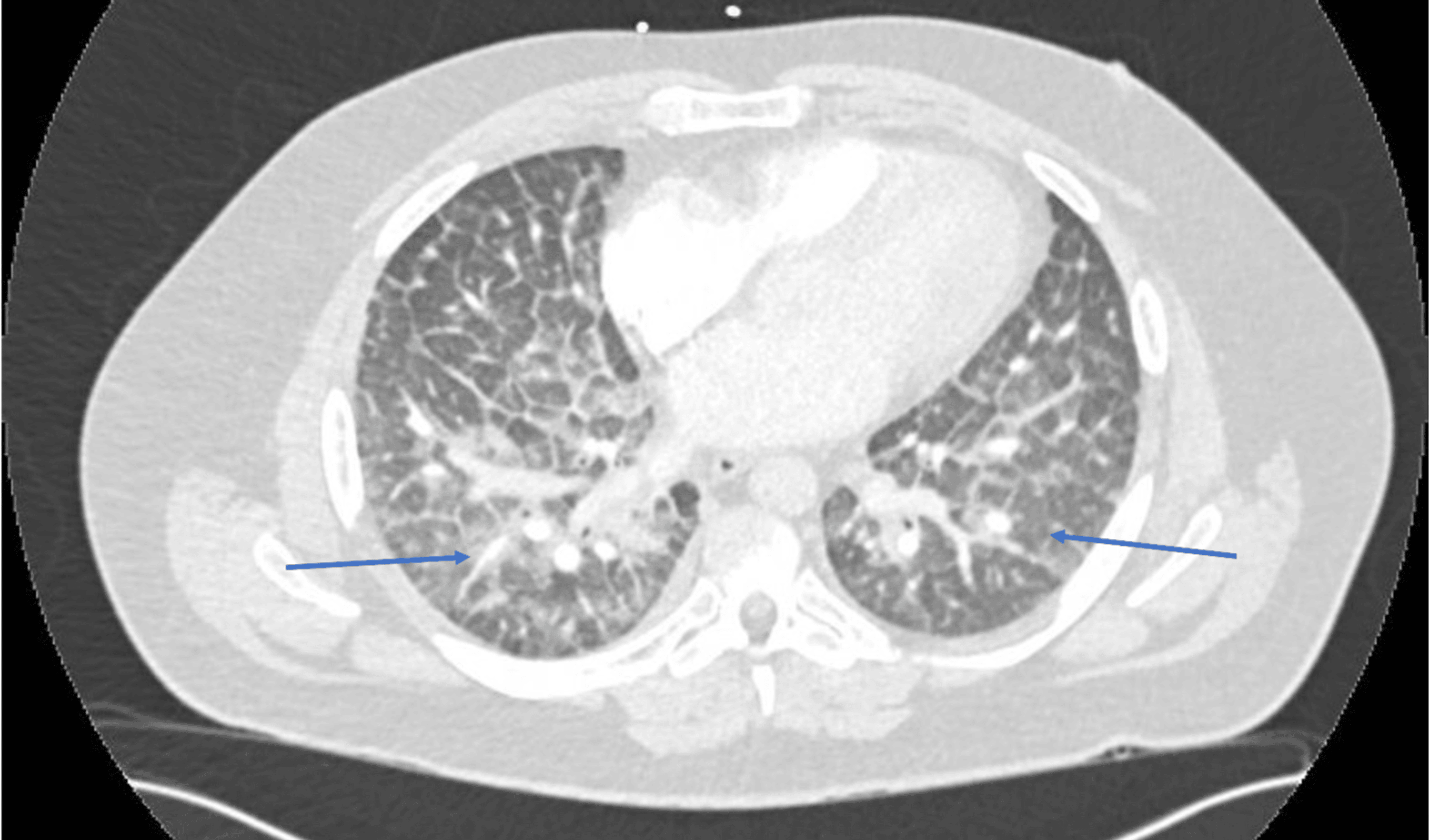
Arterial blood gas analysis revealed type 1 respiratory failure and an elevated lactate of 5 mmol/L (see Table 2). Computed tomographic pulmonary angiography excluded pulmonary embolism but demonstrated bilateral ground-glass opacities and mild pleural effusions (Figure 3).
Due to the progressive respiratory failure and haemodynamic instability (Table 1), he was transferred to the Intensive Treatment Unit (ITU) for advanced respiratory and circulatory support.
On day three of admission, the patient’s clinical condition deteriorated, with progressive respiratory failure and haemodynamic instability. His systolic blood pressure fell to 65 mmHg, with a heart rate of 80 beats/min and an elevated lactate of 4 mmol/L. He required increasing inotropic and vasopressor support, including noradrenaline, and was transferred to the ITU, where he was intubated and mechanically ventilated with an inspired oxygen fraction (FiO₂) of 80%.
Complete heart block persisted, and repeat blood cultures were obtained, with plans for further cardiac evaluation with transthoracic echocardiography and cardiac magnetic resonance imaging. The differential diagnosis at this point was revised to include viral encephalitis complicated by viral myocarditis and possible Lyme disease.
Blood cultures subsequently isolated MSSA in two sets, raising concern for an underlying bacterial infection.
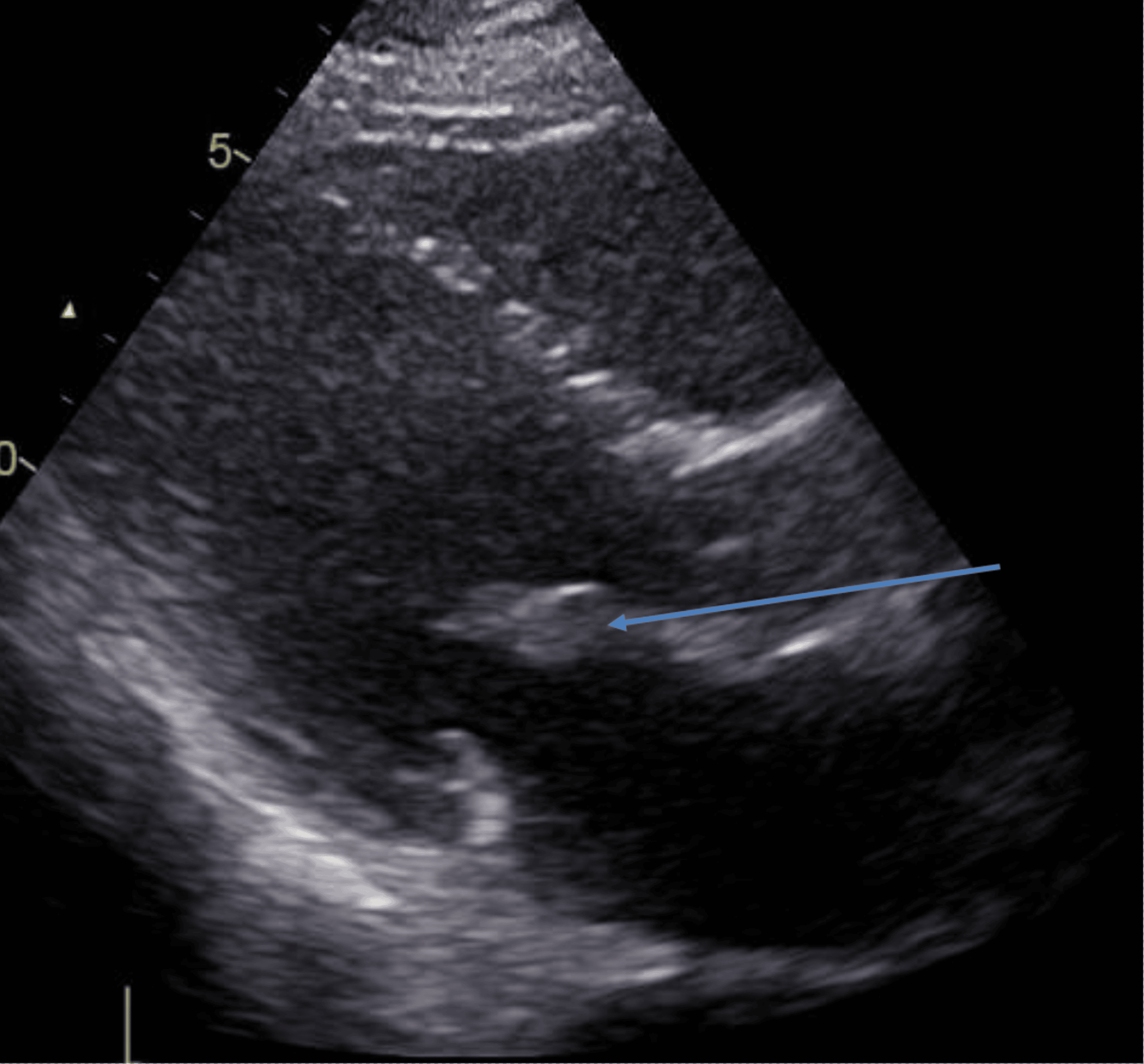
On day four of admission, bedside transthoracic echocardiography (TTE) was performed. Although the examination was technically limited, it demonstrated a mobile mass involving the anterior mitral valve leaflet, suspicious for vegetation, together with severe mitral regurgitation (Figure 4). Left ventricular systolic function was hyperdynamic.
Urgent transoesophageal echocardiography (TOE) was undertaken approximately two hours later and confirmed extensive multivalvular IE. A large vegetation was identified on the anterior mitral valve leaflet, with associated leaflet perforation and severe eccentric mitral regurgitation (Figures 5, 6). The effective regurgitant orifice area (EROA) exceeded 0.6 cm². Additional vegetations were identified on the septal leaflet of the tricuspid valve, associated with mild regurgitation, and on the pulmonary valve, without significant regurgitation. The imaging findings are summarised in Table 3.
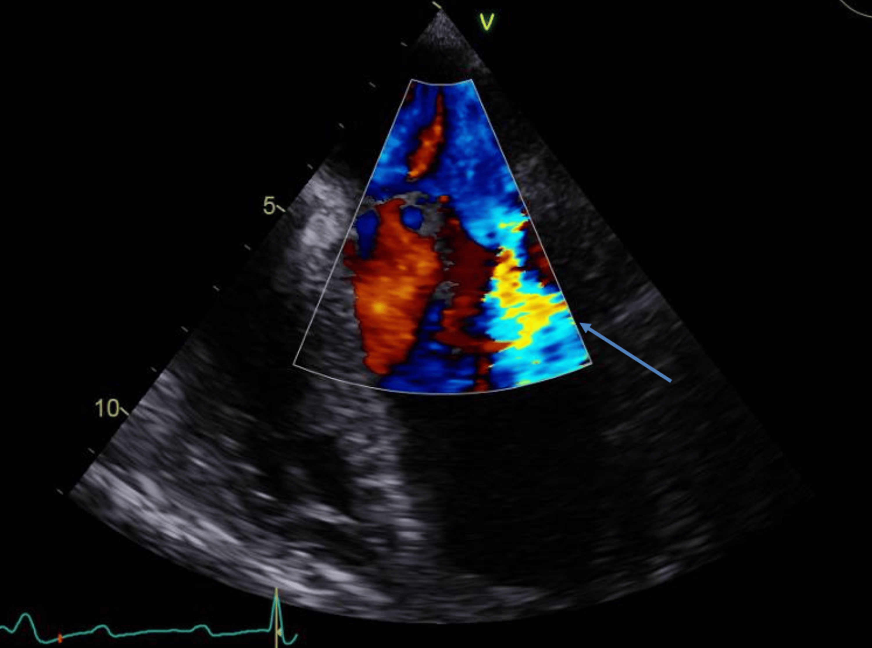
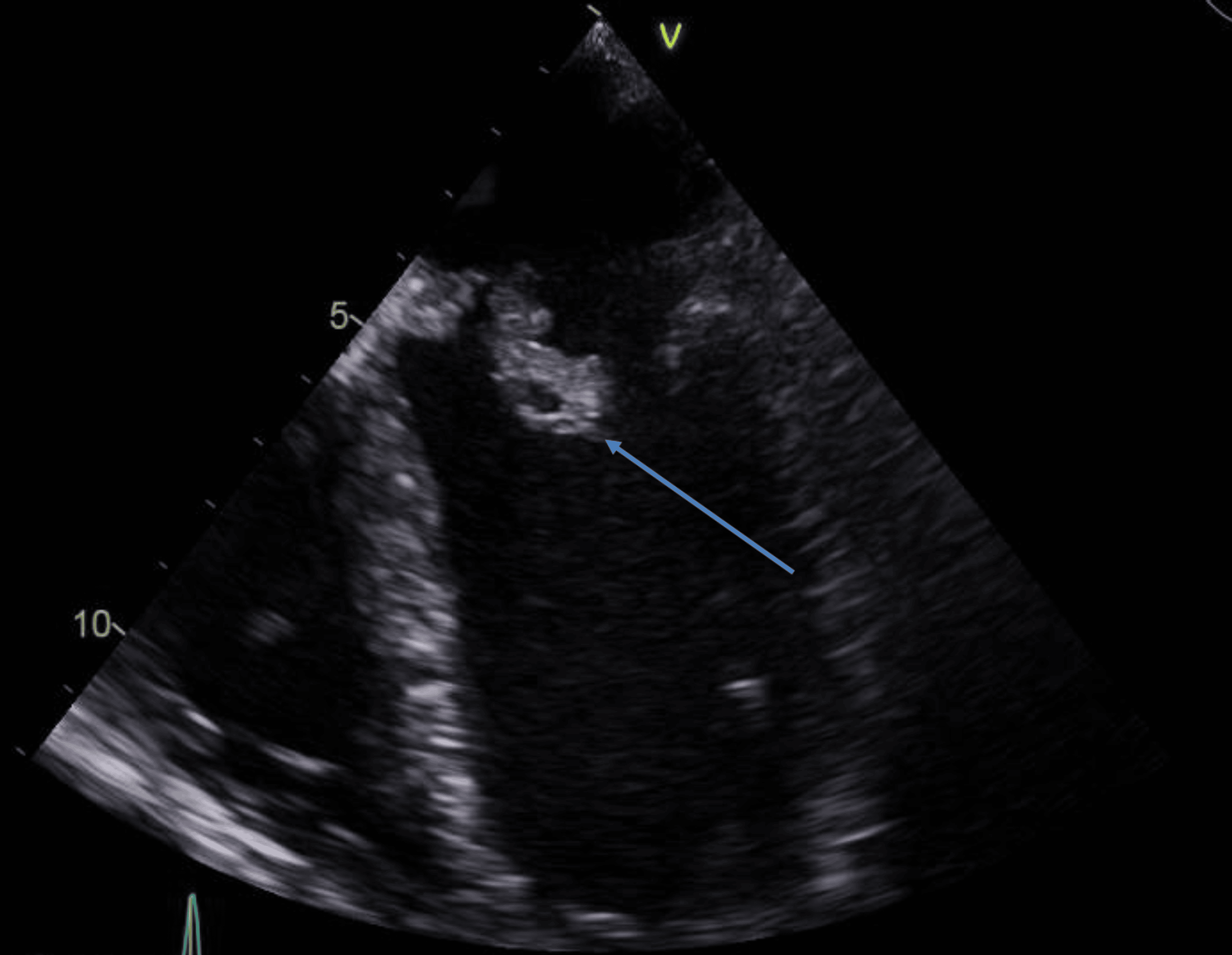
The presence of complete heart block raised concerns for peri-annular or septal extension of infection affecting the conduction system, despite the absence of a definitive aortic root abscess seen on pre-operative imaging.
Transoesophageal echocardiography confirmed multivalvular IE, with a large mitral valve vegetation complicated by anterior leaflet perforation causing severe mitral regurgitation (see Tables 3, 4). Additional vegetations were identified on the tricuspid valve septal leaflet with mild tricuspid regurgitation and on the pulmonary valve without regurgitation. Although there was no clear evidence of an aortic root abscess, the combination of multivalvular infection and persistent complete heart block raised concern for extension, involving the cardiac conduction system.
The combination of persistent complete heart block and extensive valvular infection raised concern for peri-annular or septal extension of infection affecting the cardiac conduction system. However, no definite aortic root abscess was identified on preoperative imaging.
In view of pulmonary oedema, hypotension despite high doses of noradrenaline and dobutamine, and a rising lactate of 5, suggesting cardiogenic shock secondary to severe mitral regurgitation, emergency cardiac surgery was undertaken approximately six hours after TOE, later that evening on day four of admission. This involved mitral valve replacement with a 27-mm mechanical prosthesis, tricuspid valve repair, and reconstruction of the mitral annulus using a pericardial patch. Surgical debridement involved the mitral annulus and interventricular septum, with patch reconstruction along the inferior interventricular septum and closure of the resulting septal and annular defects.
The microbiology team recommended a total six-week course of antimicrobial therapy for MSSA IE.
On postoperative day five, the patient developed worsening dyspnoea, chest pain, fever, and confusion, accompanied by persistently elevated inflammatory markers. Chest radiography demonstrated changes suggestive of pneumonia and/or pulmonary congestion. Repeat TOE raised concern for a possible paravalvular leak involving the mechanical mitral prosthesis, with moderate-to-severe regurgitation initially reported. Following multidisciplinary discussion, conservative management with serial imaging was agreed upon because the patient remained clinically stable. Subsequent echocardiography demonstrated improvement in regurgitation severity to mild.
Complete heart block persisted following surgery, and a dual-chamber permanent pacemaker was implanted two weeks postoperatively (see Table 5). The patient subsequently made a satisfactory clinical recovery and was discharged home with planned cardiology follow-up.
Discussion
This case illustrates an unusual and diagnostically challenging presentation of IE, initially mimicking meningoencephalitis (supported by a raised white cell count in the CSF, which can occur in systemic infections or an inflammatory response). Neurological manifestations occur in approximately one-third of patients with IE and may precede the development of classical cardiac findings [1]. Such presentations may result from septic embolisation, immune-mediated phenomena, or the systemic inflammatory response associated with severe infection, making early diagnosis particularly challenging and highlighting the importance of considering IE in patients presenting with neurological symptoms and systemic features of infection [2]. The diverse clinical manifestations of IE can contribute to diagnostic challenges, and the condition remains associated with significant morbidity and mortality despite improvements in diagnosis and management [3].
Complete heart block is an uncommon but important complication of IE to consider. The development of new atrioventricular conduction abnormalities should immediately prompt consideration of peri-annular extension of infection, including abscess formation or inflammatory involvement of the cardiac conduction system. Such complications are associated with increased mortality and frequently necessitate urgent surgical intervention [4]. Although no definite peri-annular abscess was identified in this patient, the persistence of complete heart block despite successful surgical intervention strongly suggests significant inflammatory involvement of the conduction system [5].
Multivalvular IE is an uncommon presentation compared with isolated single-valve involvement but is associated with a significantly higher number of complications, including heart failure, systemic embolic events, and the requirement for surgical intervention [3]. In this case, infection affecting the mitral, tricuspid, and pulmonary valves, combined with perforation of the anterior mitral leaflet causing acute severe mitral regurgitation, led to rapid deterioration with cardiogenic shock, necessitating emergency surgery.
The current European Society of Cardiology guidelines recommend prompt transthoracic echocardiography in patients with suspected IE, with transoesophageal echocardiography indicated when complications are suspected, prosthetic material is present, or transthoracic imaging does not provide sufficient diagnostic information [1]. In this patient, initial bedside transthoracic echocardiography successfully identified mitral valve vegetation, while subsequent urgent transoesophageal imaging revealed the extent of multivalvular involvement, confirmed mitral leaflet perforation, and clarified the mechanism of acute mitral regurgitation, providing key information for surgical planning.
Early involvement of a multidisciplinary team, including emergency medicine, intensive care, cardiology, microbiology, cardiac imaging, and cardiothoracic surgery, was essential in achieving a favourable outcome [6]. This case highlights the importance of prompt recognition of IE in patients presenting with systemic infection and unexplained conduction abnormalities, enabling timely initiation of antimicrobial therapy, advanced imaging, and early surgical assessment in line with current guideline recommendations.
A further educational lesson from this case relates to the importance of postoperative management and decision-making. Early postoperative transoesophageal echocardiography identified severe paravalvular mitral regurgitation, which initially raised concern for potential surgical failure or the need for repeat intervention. However, given the patient’s clinical stability and reassuring observations, a conservative approach was adopted following multidisciplinary team discussion. Subsequent echocardiography demonstrated progressive improvement, with only mild residual paravalvular regurgitation, a well-seated mitral valve prosthesis and preserved left ventricular systolic function. This case provides an important insight that, in appropriately selected stable patients, careful clinical assessment combined with serial echocardiographic monitoring may avoid unnecessary repeat surgery.
Finally, despite requiring complex reconstructive cardiac surgery, including mitral valve replacement, pericardial patch repair of the annulus, interventricular septal repair, tricuspid valve repair, and permanent dual-chamber pacemaker implantation, the patient achieved an excellent clinical recovery.
Follow-up demonstrated preserved left ventricular systolic function, only mild residual mitral regurgitation, stable tricuspid valve repair, and good functional status.
Conclusions
IE may initially present with neurological symptoms and systemic infection, delaying recognition of the underlying cardiac pathology. This case highlights the importance of considering IE in patients presenting with fever, neurological symptoms, and unexplained conduction abnormalities. New complete heart block should raise suspicion of possible peri-annular extension of infection and prompt urgent echocardiographic assessment, although the underlying mechanism may not always be confirmed. Early recognition of severe valvular complications, including leaflet perforation and acute severe regurgitation, is essential. Prompt multidisciplinary involvement and timely surgical intervention are particularly important in patients with haemodynamic deterioration. Long-term follow-up is required to assess prosthetic valve function, pacemaker status, and recurrence of infection.
References
- Delgado V, Ajmone Marsan N, de Waha S, et al.: 2023 ESC guidelines for the management of endocarditis. Eur Heart J. 2023, 44:3948-4042. 10.1093/eurheartj/ehad193
- Holland TL, Baddour LM, Bayer AS, Hoen B, Miro JM, Fowler VG Jr: Infective endocarditis. Nat Rev Dis Primers. 2016, 2:16059. 10.1038/nrdp.2016.59
- Kyriakoulis I, Tzoumas A, Kyriakoulis KG, et al.: Multivalvular vs single-valve infective endocarditis: a systematic review and meta-analysis. Future Cardiol. 2025, 21:113-21. 10.1080/14796678.2025.2457898
- Murdoch DR, Corey GR, Hoen B, et al.: Clinical presentation, etiology, and outcome of infective endocarditis in the 21st century: the International Collaboration on Endocarditis-Prospective Cohort Study. Arch Intern Med. 2009, 169:463-73. 10.1001/archinternmed.2008.603
- Infective Endocarditis Cahill TJ, Prendergast BD: Infective endocarditis. Lancet. 2016, 387:882-93. 10.1016/S0140-6736(15)00067-7
- Baddour LM, Wilson WR, Bayer AS, et al.: Infective endocarditis in adults: diagnosis, antimicrobial therapy, and management of complications: a scientific statement for healthcare professionals from the American Heart Association. Circulation. 2015, 132:1435-86.