
Anesthesiology News
University of Utah Health
Salt Lake City, Utah
University of Utah Health
Salt Lake City, Utah
University of Utah Health
Salt Lake City, Utah
University of Utah Health
Salt Lake City, Utah
University of Utah Health
Salt Lake City, Utah
Introduction
Osteogenesis imperfecta (OI) is a congenital connective tissue disease characterized by increased bone fragility, low bone mass, and other connective tissue manifestations.1 It has an incidence of 1 in 20,000 to 1 in 60,000. The disease results from mutations in the gene encoding type 1 collagen, which is the major collagen comprising bones and other connective tissues.2There is a wide variety of clinical severity in OI. In addition to increased bone fragility, there can be other defects, including cardiovascular abnormalities and coagulation abnormalities.3
There are 8 different types of OI. The most common form is OI type I, which is inherited in an autosomal dominant fashion and is characterized by blue sclera, joint hypermobility, and multiple fractures prior to puberty.4 OI type III is inherited in both dominant and recessive patterns, and it can also occur with de novo mutations. Affected patients may have a normal birth weight and length; however, multiple fractures lead to progressive deformity in the first 2 decades of life.5
Pregnancy in women who have OI is rare and carries a high risk for maternal morbidity. These pregnancies should be managed by a multidisciplinary team and patients need to be aware of the risks, so they can balance them against the benefit of having a child. The mode of delivery often depends on obstetric indications.6 However, because of the fragility of the maternal skeleton and cranio-pelvic mismatch, cesarean delivery is often preferred.9
Written HIPAA authorization was obtained from the patient in this report, and the document adheres to the applicable EQUATOR (CARE) guidelines.
Clinical Scenario
A 26-year-old woman, gravida 3, para 2, was admitted from clinic at a gestation of 31 weeks and 5 days for a 1-week history of cough and worsening shortness of breath (Figures 1-3). At admission, the patient’s weight was 34.9 kg and height was 34 inches. She had a past medical history of de novo OI type III, severe scoliosis, gastroesophageal reflux disease, restrictive lung disease, and mild intermittent asthma, with a self-reported history of more than 300 fractures. At admission, a respiratory panel was negative and she was treated conservatively.
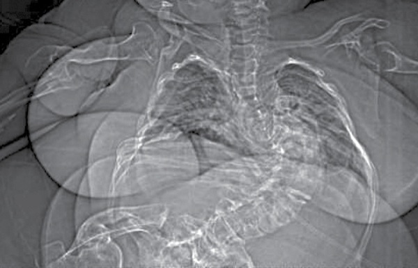
Figure 1.
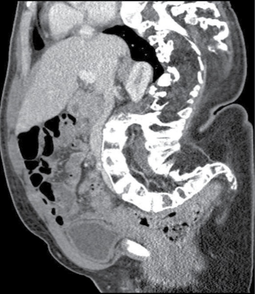
Figure 2.

Figure 3.
During the patient’s hospitalization, she was diagnosed with gestational diabetes and pre-eclampsia without severe features. After discussion with the obstetric team, a plan was made for expectant inpatient management with cesarean delivery and bilateral tubal ligation to be performed at 33 weeks’ gestation.
At a gestation of 32 weeks and 5 days, the patient began to experience increased blood pressure. She was diagnosed with gestational diabetes and pre-eclampsia that advanced to early hemolysis, elevated liver enzymes, low platelet count (HELLP) syndrome. The concern about early HELLP syndrome prompted the decision to proceed urgently with a cesarean delivery.
The patient had a Mallampati class I airway, limited cervical neck range of motion, and a thyromental distance of less than 2 cm. Preoperatively, she received 30 mL of sodium citrate. She was positioned supine on the operating table, with a roll under her right hip and careful placement of padding and blankets to minimize the risk for fracture. Standard monitors were placed. To avoid excessive neck extension and rapid desaturation as a result of her limited functional residual capacity, the patient underwent an awake fiber-optic intubation. She was also given 3 L of oxygen by nasal cannula, 0.2 mg of glycopyrrolate, and 5% lidocaine jelly to topicalize her upper airway; in addition, her posterior oropharynx and vocal cords were sprayed with nebulized 2% lidocaine. A 5.0 cuffed endotracheal tube was able to be advanced into the trachea, with 20 mg of propofol supplemented during endotracheal intubation.
After intubation, propofol was supplemented and the patient was maintained on desflurane. Following a successful cesarean delivery, she was supplemented with fentanyl and maintained on 0.5 minimum alveolar concentration (MAC) of nitrous oxide and 0.5 MAC of desflurane. Given the patient’s small stature, the oxytocin infusion was started at 9 units per hour, which was half of our institution’s standard starting dose.
Throughout the case, the patient was ventilated with pressure-controlled ventilation. To achieve a tidal volume of 6 mL/kg, peak pressures were 31 cm H2O before delivery and 21 cm H2O after delivery.
Prior to extubation, the patient was given 100 mcg of fentanyl and 0.2 mg of hydromorphone. She was also given 20 mg of furosemide to avoid pulmonary edema from placental autotransfusion, 12 puffs of albuterol for her history of asthma, and nebulized endotracheal lidocaine. She was successfully extubated. During the procedure, the patient received 800 mL of lactated Ringer’s; estimated blood loss was 500 mL; and urine output was 125 mL.
The patient’s postoperative pain was controlled with an additional 250 mcg of fentanyl, 500 mg of IV acetaminophen, and hydromorphone patient-controlled analgesia that was titrated to 0.15 mg every 10 minutes with no background infusion. She did require bilevel positive airway pressure (BiPAP) for a short time postoperatively.
The postoperative course was complicated by a wound infection that was treated with IV antibiotics. She was successfully discharged on postoperative day 14. There were no signs of OI in her child; however, no genetic testing was done.
Discussion
As discussed above, depending on the inheritance pattern of OI, disease severity can vary significantly. OI superimposed on the preexisting physiologic changes of pregnancy presents unique anesthetic challenges (Table).7
| Table. Unique Anesthetic Considerations for OI in Pregnancy | |
| Considerations | Specific Risks |
|---|---|
| Neuraxial anesthesia | Spinal deformity:
|
| Airway management for general anesthesia |
|
| Pulmonary physiology |
|
| Cardiac physiology |
|
| Fracture risk |
|
| Postpartum hemorrhage |
|
| Postoperative pain control |
|
| NSAIDs, nonsteroidal anti-inflammatory drugs; OI, osteogenesis imperfecta | |
Multiple physiologic changes can potentially exist with OI. Many patients have restrictive lung physiology secondary to thoracic dysmorphology.8 They are also at risk for congenital heart defects and malformation of the great vessels.8 Type I collagen makes up approximately 85% of cardiac muscle and provides stiffness in the ventricular wall. Reports of aortic rupture, left ventricular rupture, and aortic or mitral valve incompetence have been documented.9 Mitral valve prolapse and aortic root dilation are the most commonly seen cardiac abnormalities.10
Our patient was known to have moderate restrictive lung disease based on pre-pregnancy lung function tests (forced vital capacity [FVC], 0.76 [69%]; forced expiratory volume in 1 second [FEV1], 0.67 [57%]; FEV1:FVC, 89%). Her preoperative echocardiogram showed a well-functioning heart.
Although spinal anesthesia was considered, we determined it would have been nearly impossible to perform, given the degree of lumbar dysmorphology (Figure 1). In addition, the patient’s limited height and spinal deformities would have made the dosing and spread of the intrathecal medications tenuous and potentially dangerous; a high spinal requiring emergent airway protection would have been extremely detrimental in her case. Therefore, the decision was made to proceed with general endotracheal anesthesia.
The patient’s restrictive physiology was aggravated during pregnancy by her gravid uterus. Because of this extremely limited functional residual capacity, we were concerned about rapid desaturation during induction and laryngoscopy (Figure 2).
Furthermore, difficult airways are common in patients with OI as a result of the described thoracic kyphoscoliosis, in addition to the risk for fracture with direct laryngoscopy, a limited neck range of motion, and the concern about atlantoaxial dislocation and basilar invagination with extension of the neck.9,10 Consequently, the decision was made to perform an awake fiber-optic intubation on our patient. Because of her small stature, careful consideration was taken in the dosing of lidocaine, so as not to exceed the maximum dose. We were able to achieve successful topicalization of the airway without exceeding the maximum dose. We also chose an awake intubation to avoid a lethal hyperkalemic response with succinylcholine during rapid sequence induction, as she was partly wheelchair-bound. In addition, case reports of contraction-induced fractures have been documented in patients with OI.9 By allowing the patient to be awake and well positioned during intubation, we also were confident that the positioning would minimize the risk for fracture from malpositioning.
Measurement of blood pressure in patients with OI can be tenuous. Some researchers recommend avoiding mechanical noninvasive blood pressure cuffs, as they have been shown to cause compression-induced fractures in these patients.9 Despite this, a noninvasive blood pressure cuff was used for our patient since admission, without harm, so this method was continued intraoperatively.
In patients with OI, the risk for postpartum hemorrhage is elevated due to uterine atony, surgical lacerations, and a well-documented impairment of platelet aggregation.11 Our patient had an increased risk because of possible early HELLP syndrome. Because of her small stature, the dosing of the post–cesarean delivery oxytocin was halved. With this dose, we were able to achieve adequate uterine tone, but we were prepared to use other measures if atony or bleeding occurred.
Because of the potential impaired platelet aggregation, in addition to the fact that our patient had early HELLP syndrome, ketorolac and other nonsteroidal anti-inflammatory drugs were avoided and postoperative pain was controlled with acetaminophen and opioids. We had a low threshold to use BiPAP while she was being treated with IV opioids postoperatively, again because an airway emergency would have been extremely detrimental in her situation.
Pregnancy in patients with OI is very rare, and the pathophysiologic changes associated with OI compounded by the physiologic changes that occur with pregnancy present extremely unique anesthetic considerations. This report has shown a case of successful perioperative management of a pregnant patient with OI type III.
References
- Rauch F, Glorieux FH. Osteogenesis imperfecta. Lancet. 2004;363(9418):1377-1385.
- Litos M, Michala S, Brown R. Osteogenesis imperfecta and pregnancy. Eur J Obstet Gynecol Reprod Biol. 2008;136(1):126-127.
- Carlson W, Harlass EF. Management of osteogenesis imperfecta in pregnancy. J Reprod Med. 1993;38(3):228-232.
- Fauci A, Kasper DL, Longo D, et al. Heritable disorders of connective tissue. In: Harrison’s Principles of Internal Medicine. 17th ed. New York, NY: McGraw-Hill; 2008.
- Sharma A, George L, Erksin K. Osteogenesis imperfecta in pregnancy: two case reports and review of literature. Obstet Gynecol Surv. 2001;56(9):563-566.
- Lyra TG, Pinto VA, Ivo FA, et al. Osteogenesis imperfecta in pregnancy: case report. Revista Brasileira de Anestesiologia. 2010;60(3):321-324.
- Hwang DK, Jong IO, Hea JY, et al. Spinal anesthesia during cesarean section in a patient with severe osteogenesis imperfecta: a case report. Korean J Anesthesiol. 2009;57:662-665.
- Heinrich S, Tzabazis A. Anesthesia recommendations for patients suffering from osteogenesis imperfecta. 2012. Orphanet J Rare Dis. www.orpha.net/?data/?patho/?Pro/?en/?Osteogenesis_EN.pdf. Accessed May 1, 2017.
- Weis SM, Emery JL, Beker KD, et al. Myocardial mechanics and collagen structure in osteogenesis imperfecta murine. Circ Res. 2000;87(8):663-669.
- Oakley L, Reece LP. Anesthetic implications for the patient with osteogenesis imperfecta. AANA J. 2010;78(1):47-53.
- Litos M, Stavroula M, Richard B. Letters to the editor – brief communication. Osteogenesis imperfecta and pregnancy. Eur J Obstet Gynecol Reprod Biol. 2008;136(1):126-133.