

Authors Tanimoto S et al
Cureus 18(10): e118405. doi:10.7759/cureus.118405
Abstract
Patients with severe tricuspid regurgitation are at risk of developing right heart failure due to excessive changes in right ventricular preload or increases in pulmonary vascular resistance, necessitating careful perioperative hemodynamic management. We report a case of anesthetic management for laparoscopic cholecystectomy in an elderly patient with torrential tricuspid regurgitation, using transesophageal echocardiography and peripheral venous pressure monitoring.
The patient was a 79-year-old woman with a history of heart failure, atrial fibrillation, and myocardial infarction. Preoperative transthoracic echocardiography showed a maximum effective regurgitant orifice area of 0.85 cm² for tricuspid regurgitation. Right ventricular systolic function was preserved, and there was no clear echocardiographic evidence of pulmonary hypertension. General anesthesia was induced and maintained with remimazolam and remifentanil, and bilateral ultrasound-guided nerve blocks were performed. Transesophageal echocardiography performed after the induction of anesthesia demonstrated a vena contracta area of 1.56 cm² on three-dimensional color Doppler imaging, consistent with torrential tricuspid regurgitation.
Intraoperatively, pneumoperitoneum pressure was gradually increased from a low level, and the patient was slowly transitioned to the head-up position. Ventilation was managed to avoid hypercapnia. Transesophageal echocardiography was used to quantitatively assess the severity of tricuspid regurgitation after the induction of anesthesia and to visually assess changes in tricuspid regurgitation before and after pneumoperitoneum and positional changes. Serial changes in peripheral venous pressure were used as an adjunctive indicator of right-sided filling pressure and systemic venous congestion. No visually apparent worsening of tricuspid regurgitation or sustained increase in peripheral venous pressure was observed following pneumoperitoneum or positional changes, and hemodynamics remained generally stable throughout the procedure. The surgery was completed without complications, and the patient was discharged on postoperative day 4.
While performing laparoscopic surgery for a patient with torrential tricuspid regurgitation, combining the transesophageal echocardiographic assessment of tricuspid regurgitation with the serial monitoring of peripheral venous pressure provided an adjunctive means of evaluating changes in right-sided hemodynamics associated with pneumoperitoneum and positional changes. Because peripheral venous pressure is not an indicator of fluid responsiveness, it should not be interpreted in isolation but rather integrated with other hemodynamic parameters, including transesophageal echocardiographic findings and arterial blood pressure.
Introduction
In patients with severe tricuspid regurgitation, excessive increases or decreases in right ventricular preload and elevations in pulmonary vascular resistance may precipitate right heart failure. Therefore, perioperative management should aim to maintain venous return while avoiding excessive right-sided volume loading and increases in right ventricular afterload [1,2]. During laparoscopic surgery, changes in venous return, airway pressure, and pulmonary vascular resistance associated with pneumoperitoneum and positional changes may affect right-sided hemodynamics [1,2]. Furthermore, Parikh et al. reported that moderate and severe tricuspid regurgitation, classified according to conventional severity grading, were independently associated with increased 30-day mortality after noncardiac surgery, irrespective of left ventricular ejection fraction and the Revised Cardiac Risk Index [3]. In recent years, a five-grade classification of tricuspid regurgitation severity comprising mild, moderate, severe, massive, and torrential grades has been proposed, with torrential tricuspid regurgitation representing the most severe category [4]. The proposed quantitative criteria for each grade are summarized in the Appendices. We report a case of an elderly patient with torrential tricuspid regurgitation who underwent laparoscopic cholecystectomy with intraoperative management combining the transesophageal echocardiographic assessment of right heart function and peripheral venous pressure monitoring. Peripheral venous pressure monitoring may provide an adjunctive means of assessing changes in right-sided filling pressure and systemic venous congestion without central venous catheterization.
Case Presentation
A 79-year-old woman with a height of 157.2 cm and a weight of 49.8 kg was scheduled to undergo laparoscopic cholecystectomy for cholelithiasis following treatment for choledocholithiasis. Her medical history included heart failure, atrial fibrillation, diabetes mellitus, and myocardial infarction. Although the details of the myocardial infarction were unavailable, she had a history of percutaneous coronary intervention. Preoperatively, she had no apparent symptoms of heart failure, and physical examination revealed no remarkable abnormalities. Her medications included rivaroxaban 10 mg/day, azosemide 15 mg/day, spironolactone 25 mg/day, bisoprolol 2.5 mg/day, and empagliflozin 10 mg/day. Rivaroxaban was discontinued on the day before the scheduled surgery.
Preoperative laboratory tests showed a blood urea nitrogen level of 23.2 mg/dL, serum creatinine level of 1.13 mg/dL, estimated glomerular filtration rate of 35.8 mL/minute/1.73 m², and brain natriuretic peptide level of 123 pg/mL. No other significant abnormalities were identified.
Chest radiography showed cardiomegaly, with a cardiothoracic ratio of 60.3%. A 12-lead electrocardiogram showed atrial fibrillation. Pulmonary function testing revealed no significant abnormalities. Transthoracic echocardiography showed a left ventricular ejection fraction of 47% with diffuse left ventricular hypokinesis. The left atrial volume was 91 mL, with a left atrial volume index of 61 mL/m², indicating marked left atrial enlargement. The right atrium was also markedly enlarged, with a longitudinal diameter of 61 mm, a transverse diameter of 56 mm, and an area of 41 cm². Tricuspid annular plane systolic excursion (TAPSE) was 18.1 mm, tissue Doppler-derived lateral tricuspid annular systolic velocity (S′) was 10.4 cm/second, and right ventricular fractional area change (FAC) was 44%, indicating preserved right ventricular systolic function. A coaptation gap of approximately 8.6 mm was observed between the tricuspid valve leaflets. Severe or greater tricuspid regurgitation was present. The proximal isovelocity surface area (PISA) radius ranged from 8.2 to 10.0 mm, and the effective regurgitant orifice area (EROA) calculated using the PISA method ranged from 0.57 to 0.85 cm². The number of cardiac cycles used for these measurements was not specified in the original echocardiography report. The mean vena contracta width measured in two imaging planes was 7.9 mm. Systolic flow reversal in the hepatic veins was also observed. The EROA varied from beat to beat, ranging from values consistent with severe to torrential tricuspid regurgitation. The maximum EROA of 0.85 cm² met the criterion for torrential tricuspid regurgitation. The inferior vena cava diameter was 20 mm, with respiratory variation of ≥50%, corresponding to an estimated right atrial pressure of 3 mmHg. The peak tricuspid regurgitant pressure gradient was 19 mmHg, yielding an estimated right ventricular systolic pressure of 22 mmHg. Echocardiography showed no clear evidence of pulmonary hypertension.
The patient was managed perioperatively as having torrential tricuspid regurgitation. To minimize hemodynamic fluctuations, remimazolam was selected for both the induction and maintenance of anesthesia. In addition, a transesophageal echocardiography probe was planned for insertion to allow the real-time assessment of changes in cardiac function associated with pneumoperitoneum and positional changes. Preoperative transthoracic echocardiography showed no clear evidence of pulmonary hypertension, and right ventricular systolic function was preserved. Furthermore, because the planned procedure was laparoscopic cholecystectomy, the likelihood of massive blood loss or the need for rapid large-volume fluid administration was considered low. Therefore, the potential benefit of central venous catheter placement was considered limited, and we decided not to insert one. Instead, a peripheral intravenous catheter was placed in an upper extremity, and peripheral venous pressure was continuously monitored as an adjunctive parameter to assess changes in venous pressure.
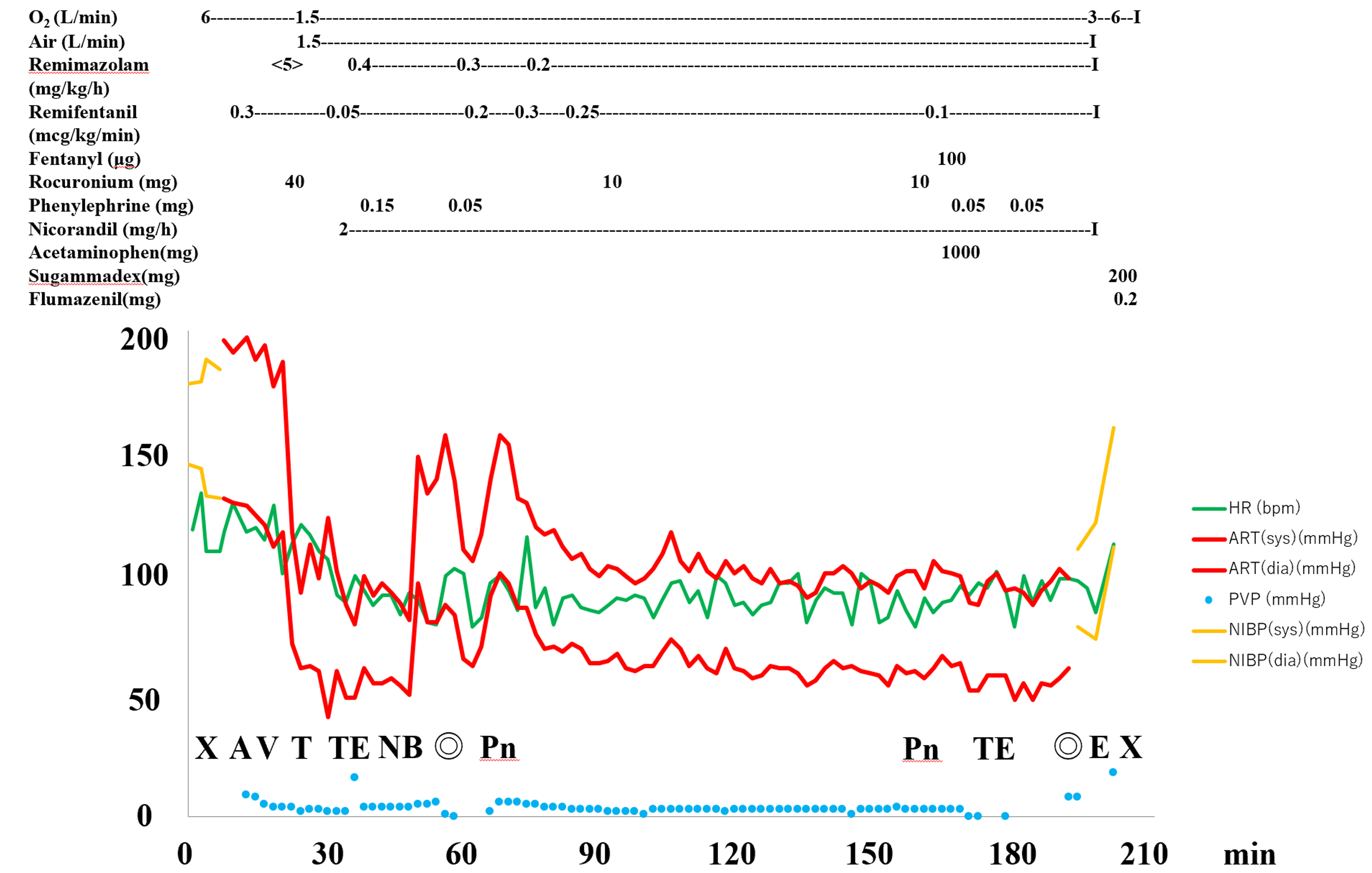
General anesthesia was performed in combination with a peripheral nerve block. Before the induction of anesthesia, a radial arterial catheter was inserted under local anesthesia. For peripheral venous pressure monitoring, a 20 G peripheral intravenous catheter (Supercath®, Medikit Co., Ltd., Tokyo, Japan) was placed in the right upper arm. The pressure transducer was zeroed at the level of the right atrium. The monitored arm was maintained at the same level as the torso, and no medications or fluids were administered through the catheter used for pressure measurement. Peripheral venous pressure was 5 mmHg before the induction of anesthesia. Immediately before pneumoperitoneum, peripheral venous pressure was 1 mmHg, which transiently increased to 6 mmHg following insufflation and further to 7 mmHg after transition to the head-up position. Subsequently, peripheral venous pressure decreased to approximately 3 mmHg during pneumoperitoneum and returned to 1 mmHg after desufflation. No sustained increase in peripheral venous pressure was observed. Anesthesia was induced with a single 5 mg dose of remimazolam, a continuous infusion of remifentanil at 0.3 μg/kg/minute, and 40 mg of rocuronium. After tracheal intubation, a transesophageal echocardiography probe was inserted.
With the patient in the supine position, bilateral space between the endothoracic fascia, diaphragm, and costodiaphragmatic recess (SEDIC) blocks were performed under ultrasound guidance using an ultrasound system (Sonosite® ST; FUJIFILM, Tokyo, Japan). A total of 40 mL of 0.25% levobupivacaine was administered for the blocks.
Anesthesia was maintained with air, oxygen, and remimazolam. Remimazolam infusion was initiated at 0.4 mg/kg/hour. Remifentanil was titrated between 0.05 and 0.30 μg/kg/minute according to the hemodynamic response and the intensity of surgical stimulation.
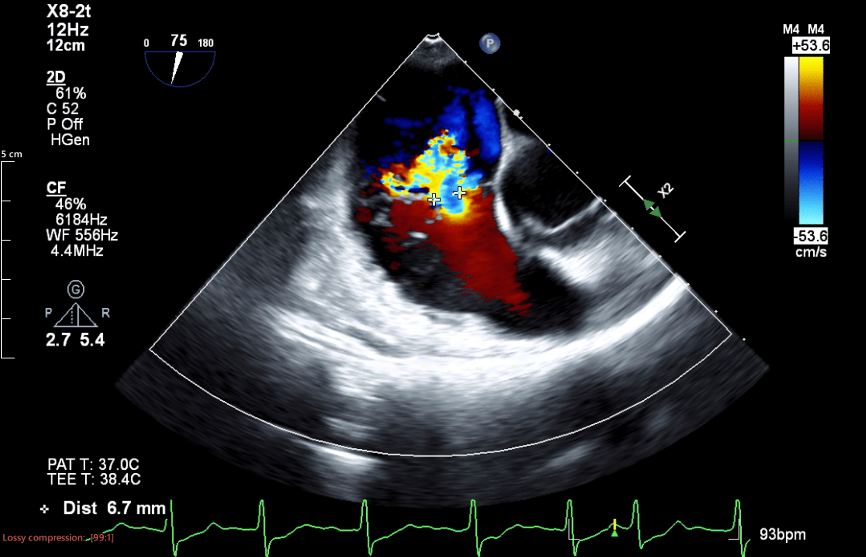
Before the start of surgery, right heart function and the severity of tricuspid regurgitation were assessed using transesophageal echocardiography. The vena contracta width measured by two-dimensional color Doppler imaging was 6.7 mm (Figure 1), and the vena contracta area measured by three-dimensional color Doppler imaging was 1.56 cm² (Figure 2).
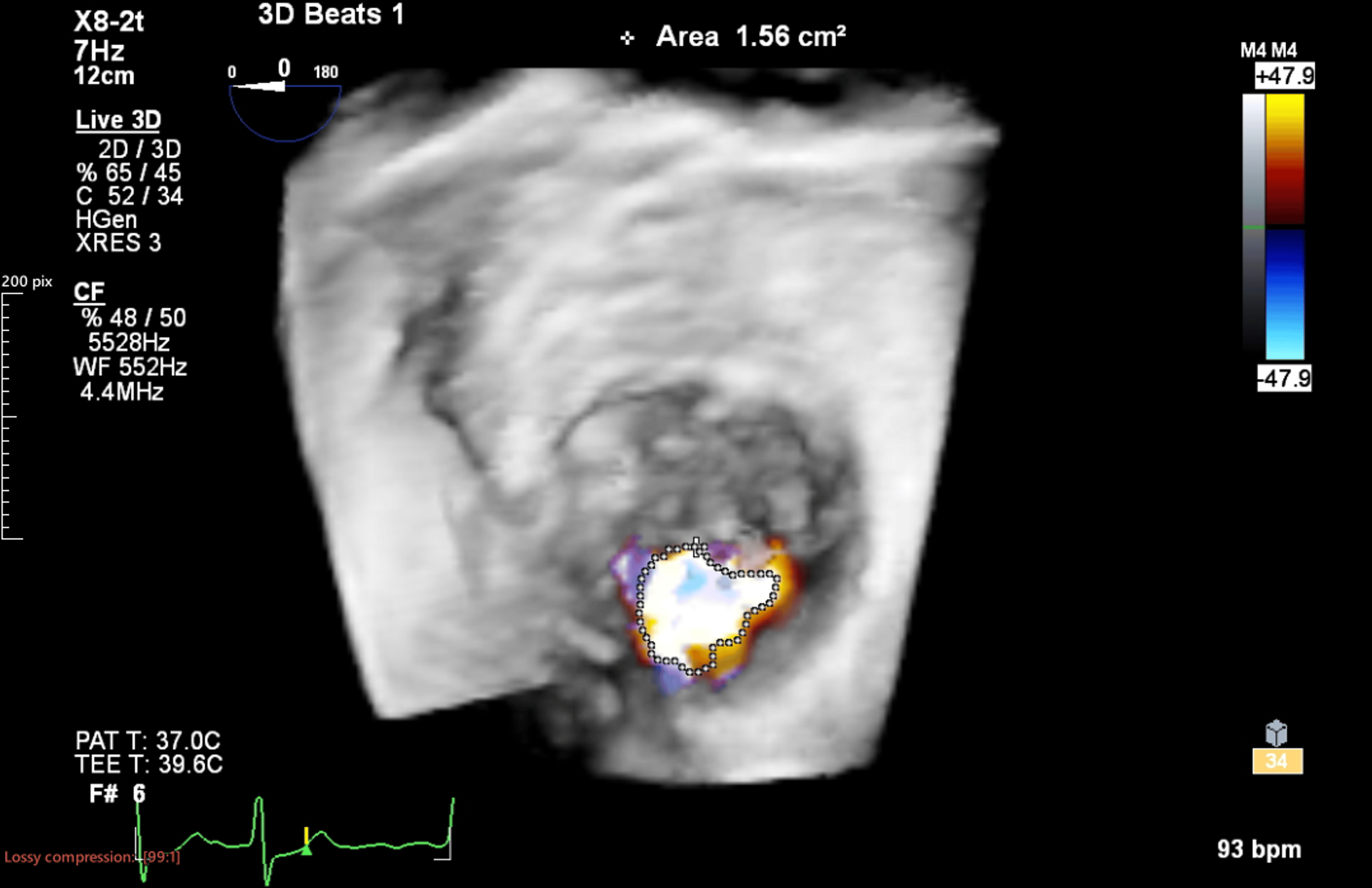
The three-dimensional vena contracta area exceeded the quantitative threshold of 1.15 cm² for torrential tricuspid regurgitation, confirming findings consistent with torrential tricuspid regurgitation on intraoperative transesophageal echocardiography. After discussion with the gastrointestinal surgeons, the pneumoperitoneum pressure was increased stepwise to a level sufficient to obtain an adequate surgical field, while hemodynamics and transesophageal echocardiographic findings were closely monitored. The transition to the head-up position was also performed gradually while hemodynamics and transesophageal echocardiographic findings were closely monitored. The exact initial and maximum pneumoperitoneum pressures and the degree of head-up positioning were not documented. Before the start of surgery, arterial blood gas analysis showed a partial pressure of carbon dioxide (PaCO₂) of 41.7 mmHg, while the corresponding end-tidal carbon dioxide (EtCO₂) was 38 mmHg. Taking this difference into account, ventilatory settings were adjusted based on continuous EtCO₂ monitoring to maintain EtCO₂ at ≤40 mmHg in an effort to minimize hypercapnia and prevent an increase in pulmonary vascular resistance. Transesophageal echocardiography was used to visually assess changes in tricuspid regurgitation before and after pneumoperitoneum and positional changes, and no apparent worsening was observed. However, quantitative measurements of right ventricular size and systolic function were not obtained. Peripheral venous pressure did not increase substantially during surgery, and hemodynamics remained stable without the need for catecholamine administration. For postoperative analgesia, 1,000 mg of acetaminophen and 50 mg of flurbiprofen were administered.
The surgery was completed without complications. The operative time was two hours and 14 minutes, and the duration of anesthesia was three hours and 26 minutes (Figure 3).
The total intraoperative fluid administration was 450 mL, urine output was 200 mL, and estimated blood loss was minimal. Postoperatively, the patient was managed in the general ward without requiring intensive cardiovascular monitoring. No clinical signs suggestive of right-sided heart failure were observed during the postoperative period. The patient was discharged on postoperative day 4 without complications. No echocardiographic examination was performed during the postoperative follow-up period, and the patient remained alive at 30 days after surgery.
Discussion
Parikh et al. reported that moderate and severe tricuspid regurgitation, classified according to conventional severity grading, were independently associated with increased 30-day mortality after noncardiac surgery, irrespective of left ventricular ejection fraction and the Revised Cardiac Risk Index [3]. Perioperative risk is further increased in the presence of right ventricular dysfunction or elevated pulmonary artery pressure [3].
In the present case, the EROA values reported on preoperative transthoracic echocardiography ranged from 0.57 to 0.85 cm², with the maximum value meeting the quantitative criterion for torrential tricuspid regurgitation of ≥0.80 cm² (see Appendices) [4]. However, the number of cardiac cycles assessed was not documented, and beat-to-beat variability associated with atrial fibrillation may have influenced the measurements. The mean vena contracta width measured in two imaging planes was 7.9 mm, corresponding to severe tricuspid regurgitation according to the proposed quantitative criteria (see Appendices). Furthermore, after the induction of anesthesia, transesophageal echocardiography showed a two-dimensional color Doppler vena contracta width of 6.7 mm, whereas the three-dimensional color Doppler vena contracta area was 1.56 cm², exceeding the quantitative criterion for torrential tricuspid regurgitation of ≥1.15 cm² [4]. Therefore, the severity of tricuspid regurgitation was assessed by integrating multiple echocardiographic parameters rather than relying solely on the maximum EROA.
Integrating these multiple echocardiographic findings, we considered that the patient had advanced tricuspid regurgitation consistent with torrential tricuspid regurgitation despite having few clinical symptoms. Right ventricular systolic function, as assessed by TAPSE, S′, and right ventricular fractional area change, was preserved, and there was no clear evidence of pulmonary hypertension. However, in patients with torrential tricuspid regurgitation, Doppler-derived pressure gradients may underestimate right ventricular systolic pressure because of rapid systolic pressure equalization between the right ventricle and right atrium. Therefore, pulmonary hypertension could not be definitively excluded based on the estimated right ventricular systolic pressure alone. Thus, although findings suggestive of chronic right-sided volume overload were present, there was no clear evidence of right ventricular systolic dysfunction or increased right ventricular afterload. In the anesthetic management of patients with severe tricuspid regurgitation, it is important to maintain venous return while avoiding excessive right-sided volume loading, prevent increases in pulmonary vascular resistance, and preserve right ventricular perfusion pressure and right ventricular contractility [1,2]. In tricuspid regurgitation, chronic right ventricular volume overload is present; therefore, excessive fluid administration may further dilate the right atrium and right ventricle, promote tricuspid annular dilatation, and increase the regurgitant volume, potentially resulting in a reduction in stroke volume. Conversely, the induction of anesthesia, positive-pressure ventilation, pneumoperitoneum, and transition to the head-up position may reduce venous return, and an excessive reduction in preload may decrease cardiac output. Therefore, preload should be carefully titrated to maintain cardiac output without exacerbating right-sided volume overload [1,2]. Furthermore, because the right ventricle has limited tolerance to increases in afterload, hypoxemia, hypercapnia, acidosis, high airway pressures, and excessive positive end-expiratory pressure should be avoided to prevent increases in pulmonary vascular resistance [1,2]. During laparoscopic surgery, increased intra-abdominal pressure caused by pneumoperitoneum may alter venous return and increase right ventricular afterload through diaphragmatic elevation and increased airway pressure.
In the present case, pneumoperitoneum pressure was increased stepwise from a low level, transition to the head-up position was performed gradually, and ventilatory settings were adjusted to avoid hypercapnia. These measures were considered physiologically appropriate for minimizing abrupt changes in right ventricular preload and afterload. In addition, because the patient had atrial fibrillation, it was important to maintain the heart rate within an appropriate range while avoiding excessive tachycardia and bradycardia, thereby preserving right ventricular filling time and cardiac output.
Remimazolam was selected for both the induction and maintenance of anesthesia with the aim of minimizing hemodynamic fluctuations. A peripheral nerve block was also used to reduce the requirements for general anesthetics and opioids. Intraoperative transesophageal echocardiography was used to quantitatively assess the severity of tricuspid regurgitation after the induction of anesthesia and to visually assess changes in tricuspid regurgitation following pneumoperitoneum and positional changes. However, quantitative measurements of right ventricular size and systolic function were not obtained before and after these interventions, which limited the objective assessment of changes in right ventricular morphology and function. Because no visually apparent worsening of tricuspid regurgitation was observed and hemodynamics, including arterial blood pressure, remained stable, we considered that aggressive volume loading was unnecessary and managed the patient without additional fluid administration or the use of catecholamines.
Peripheral venous pressure can reflect changes in central venous pressure or right atrial pressure when a continuous hydrostatic column of blood is maintained between the peripheral veins of the upper extremity and the central venous system, provided that there is no obstruction or external compression along the measurement pathway. In a study of 150 surgical patients, peripheral venous pressure was approximately 2 mmHg higher than central venous pressure on average; however, a strong correlation was observed between the two measurements, suggesting that central venous pressure may be estimated from peripheral venous pressure with reasonable accuracy [5]. Similarly, a study analyzing 1,026 paired measurements obtained from 15 neurosurgical patients demonstrated a good correlation between temporal changes in peripheral venous pressure and central venous pressure, with particularly strong correlations observed in patients with greater blood loss or larger hemodynamic fluctuations [6]. Furthermore, a strong correlation between peripheral venous pressure and central venous pressure has also been demonstrated during the rapid hemodynamic changes encountered in liver transplantation [7].
Regarding catheter placement, studies have shown no substantial difference in agreement with central venous pressure when peripheral venous pressure is measured from either an antecubital vein or a dorsal hand vein [8]. In patients without cardiac disease undergoing laparoscopic colorectal surgery, peripheral venous pressure showed a strong correlation and acceptable agreement with central venous pressure even during changes in intrathoracic pressure associated with pneumoperitoneum and the Trendelenburg position [9]. Although the patient population and surgical position differed from those in the present case, these findings support the feasibility of the trend monitoring of peripheral venous pressure during laparoscopic surgery.
The relationship between peripheral venous pressure and right atrial pressure has also been investigated in patients with heart failure. In a study of patients with acute decompensated heart failure, the correlation coefficient between peripheral venous pressure and central venous pressure was 0.947, with a mean difference of 0.4 mmHg between the two measurements [10]. A strong correlation between peripheral venous pressure and central venous pressure has also been demonstrated in patients with heart failure, heart transplant recipients, and patients with left ventricular assist devices [11]. Furthermore, peripheral venous pressure has been shown to correlate strongly with right atrial pressure and to have greater diagnostic ability for distinguishing low and high right atrial pressures than peripheral edema, jugular venous pressure, B-type natriuretic peptide, or inferior vena cava diameter [12]. These findings suggest that peripheral venous pressure may also be useful for assessing right atrial pressure and systemic venous congestion in patients with right-sided volume overload.
However, there are important limitations to the interpretation of peripheral venous pressure. A correlation between peripheral venous pressure and central venous pressure does not imply that peripheral venous pressure can predict fluid responsiveness. The pooled area under the receiver operating characteristic curve for central venous pressure in predicting fluid responsiveness has been reported to be only 0.56, and the correlation coefficient between central venous pressure and changes in stroke volume after fluid loading was only 0.18, indicating that central venous pressure alone is not an appropriate basis for guiding fluid administration [13]. Therefore, peripheral venous pressure, which may reflect changes in central venous pressure, should likewise not be used to guide fluid loading based solely on its absolute value.
Furthermore, in patients with severe tricuspid regurgitation, elevations in right atrial pressure and peripheral venous pressure may reflect an increase in mean right atrial pressure due to systolic regurgitation and systemic venous congestion rather than an increase in preload contributing to forward stroke volume. Therefore, peripheral venous pressure should not be regarded as a direct measure of right ventricular preload or of an increase in cardiac output in response to fluid administration. Rather, it should be considered an adjunctive parameter for monitoring temporal changes in right atrial pressure and systemic venous congestion.
In the present case, peripheral venous pressure increased transiently following pneumoperitoneum and transition to the head-up position but subsequently decreased despite continued pneumoperitoneum, without a sustained elevation. Qualitative transesophageal echocardiographic assessment revealed no visually apparent progression of right ventricular dilatation or worsening of tricuspid regurgitation, and hemodynamics, including arterial blood pressure, remained stable. Taken together, these findings suggest that the stability of peripheral venous pressure did not indicate preserved fluid responsiveness but rather reflected the absence of findings suggestive of an abrupt increase in right-sided filling pressure or the progression of systemic venous congestion associated with pneumoperitoneum and positional changes. By focusing on temporal changes in peripheral venous pressure rather than its absolute value and interpreting these changes together with transesophageal echocardiographic findings and arterial blood pressure, we were able to monitor changes in right-sided filling pressure and systemic venous congestion as adjunctive hemodynamic indicators without the placement of a central venous catheter. This integrated assessment may have provided useful information for avoiding excessive fluid administration.
Conclusions
In this case of laparoscopic surgery in a patient with torrential tricuspid regurgitation, pneumoperitoneum pressure and patient positioning were adjusted gradually, and ventilation was managed to avoid increases in pulmonary vascular resistance. By combining the transesophageal echocardiographic assessment of tricuspid regurgitation with the serial monitoring of peripheral venous pressure, we were able to adjunctively monitor changes in right-sided hemodynamics associated with pneumoperitoneum and positional changes. Peripheral venous pressure monitoring may have provided useful adjunctive information in this case; however, its broader clinical utility cannot be determined from a single case, and it should not be interpreted as an indicator of fluid responsiveness or used in isolation.
References
- Arora RC, Brown JK, Chatterjee S, Gan TJ, Singh G, Tong MZ: Perioperative management of the vulnerable and failing right ventricle. Perioper Med (Lond). 2024, 13:40. 10.1186/s13741-024-00397-5
- O Ibekwe S, Deschamps J, Grocott MP, Liang Y, Shaw A, E Perry T: Perioperative Quality Initiative (POQI) consensus statement on perioperative assessment of right ventricular function. Perioper Med (Lond). 2023, 12:66. 10.1186/s13741-023-00351-x
- Parikh P, Banerjee K, Ali A, et al.: Impact of tricuspid regurgitation on postoperative outcomes after non-cardiac surgeries. Open Heart. 2020, 7:e001183. 10.1136/openhrt-2019-001183
- Hahn RT, Thomas JD, Khalique OK, Cavalcante JL, Praz F, Zoghbi WA: Imaging assessment of tricuspid regurgitation severity. JACC Cardiovasc Imaging. 2019, 12:469-90. 10.1016/j.jcmg.2018.07.033
- Amar D, Melendez JA, Zhang H, Dobres C, Leung DH, Padilla RE: Correlation of peripheral venous pressure and central venous pressure in surgical patients. J Cardiothorac Vasc Anesth. 2001, 15:40-3. 10.1053/jcan.2001.20271
- Munis JR, Bhatia S, Lozada LJ: Peripheral venous pressure as a hemodynamic variable in neurosurgical patients. Anesth Analg. 2001, 92:172-9. 10.1097/00000539-200101000-00033
- Hoftman N, Braunfeld M, Hoftman G, Mahajan A: Peripheral venous pressure as a predictor of central venous pressure during orthotopic liver transplantation. J Clin Anesth. 2006, 18:251-5. 10.1016/j.jclinane.2005.09.031
- Sahin A, Salman MA, Salman AE, Aypar U: Effect of catheter site on the agreement of peripheral and central venous pressure measurements in neurosurgical patients. J Clin Anesth. 2005, 17:348-52. 10.1016/j.jclinane.2004.08.015
- Kim SH, Park SY, Cui J, et al.: Peripheral venous pressure as an alternative to central venous pressure in patients undergoing laparoscopic colorectal surgery. Br J Anaesth. 2011, 106:305-11. 10.1093/bja/aeq399
- Sperry BW, Campbell J, Yanavitski M, Kapadia S, Tang WH, Hanna M: Peripheral venous pressure measurements in patients with acute decompensated heart failure (PVP-HF). Circ Heart Fail. 2017, 10:e004130. 10.1161/CIRCHEARTFAILURE.117.004130
- Vlismas PP, Wiesenfeld E, Oh KT, et al.: Relation of peripheral venous pressure to central venous pressure in patients with heart failure, heart transplant, and left ventricular assist device. Am J Cardiol. 2021, 138:80-4. 10.1016/j.amjcard.2020.09.055
- Maruichi-Kawakami S, Nagao K, Aida K, et al.: Peripheral venous pressure measurements to evaluate congestion in heart failure. J Card Fail. 2023, 29:1319-23. 10.1016/j.cardfail.2021.11.018
- Marik PE, Cavallazzi R: Does the central venous pressure predict fluid responsiveness? An updated meta-analysis and a plea for some common sense. Crit Care Med. 2013, 41:1774-81. 10.1097/CCM.0b013e31828a25fd
Appendices
Table 1 shows the proposed quantitative criteria for the five-grade classification of tricuspid regurgitation severity.