

Authors: Gupta M, Farag A
Cureus 18(7): e112686. doi:10.7759/cureus.112686
Abstract
This case describes a 40-year-old woman with gestational trophoblastic disease (GTD) who presented with vaginal bleeding, iron deficiency anemia, and a markedly elevated beta-human chorionic gonadotropin (β-hCG) of 1,889,800 mIU/mL. Transabdominal ultrasound revealed retained products of conception and GTD. The patient had experienced a failed pregnancy two months prior to presentation. She required a massive transfusion (five units of packed red blood cells) and underwent dilation and curettage under general anesthesia with rapid sequence intubation. This case examines the anesthetic considerations and perioperative complications associated with this high-risk case.
Introduction
Gestational trophoblastic disease (GTD) encompasses a spectrum of pregnancy-related disorders arising from the abnormal proliferation of trophoblastic tissue, ranging from benign hydatidiform moles to malignant gestational trophoblastic neoplasia [1]. GTD occurs in approximately 1-2 per 1,000 pregnancies in North America and other developed nations, with hydatidiform mole representing the most common presentation; patients classically present with vaginal bleeding, markedly elevated beta-human chorionic gonadotropin (β-hCG) for gestational age, and, in more advanced cases, pelvic pain or cramping, though earlier diagnosis via routine ultrasound and β-hCG screening has made classic presentations less common [1]. These conditions are characterized by markedly elevated β-hCG levels and carry a significant risk for hemorrhagic and endocrine complications that pose unique challenges for anesthesia providers. Massive hemorrhage during uterine evacuation can precipitate disseminated intravascular coagulation (DIC), a consumptive coagulopathy associated with up to 25% of maternal death [2]. Additionally, the structural homology between β-hCG and thyroid-stimulating hormone (TSH) enables the direct stimulation of TSH receptors, placing patients with extremely elevated β-hCG levels at risk for thyrotoxicosis and life-threatening thyroid storm [3]. Anesthetic management in this population is particularly challenging because the two dominant risks, hemorrhagic coagulopathy and thyroid storm which can present simultaneously, mimic one another and require opposing physiologic priorities including aggressive volume and blood product resuscitation versus careful hemodynamic control of a hyper-dynamic thyrotoxic state, often with little advance warning. The convergence of these complications demands meticulous preoperative assessment, vigilant intraoperative monitoring, and a multidisciplinary approach to perioperative care. Extreme β-hCG elevations sufficient to provoke both massive hemorrhage and thyroid storm in the same patient are rare, and reports detailing the anesthetic approach to this specific combination are limited. We present this case to guide anesthesiologists in recognizing and managing this uncommon but high-acuity presentation. This case report describes the anesthetic management of a 40-year-old woman with GTD complicated by iron deficiency anemia, massive transfusion, and a β-hCG level of 1,889,800 mIU/mL, highlighting the critical considerations for safe anesthetic care in this high-risk population.
Case Presentation
This is the case of a gravida 10 para 3 (G10P3073), 40-year-old female patient who presented to St. John’s Episcopal Hospital with heavy vaginal bleeding and syncope. She has a history of three term deliveries, zero preterm deliveries, and seven abortions and has three living children. The patient stated having two menstrual cycles within a month and episodes of spotting. A massive transfusion protocol (MTP) was called as the patient had lost 450 cc of blood at the bedside on presentation.
The patient has a past medical history (PMHx) of hypothyroidism, iron deficiency anemia, multiple failed pregnancies, human papillomavirus (HPV), obesity, and anxiety. Further gynecologic history provided by the patient included a Pap smear a year ago, which showed atypical squamous cells of undetermined significance (ASCUS). Additionally, the patient’s obstetrics history revealed three normal spontaneous vaginal deliveries (NSVD) and six spontaneous abortions (SAB), the most recent one being two months before presentation to the hospital. An ultrasound at the time revealed an irregular gestation sac with an 8 mm fetal pole, but no fetal heart rate was identified. The patient was asked to follow up in the clinic, but was a no-show. The patient’s current medications included levothyroxine 180 mg, iron, and prenatal vitamins. On initial physical exam, the patient’s vitals signaled hemodynamic stability, while elevated blood pressure of 142/90 raised concerns. The genitourinary female physical exam revealed normal external genitalia, and minimal blood clots were evaluated from the vaginal canal. The initial laboratory findings are summarized in Table 1.
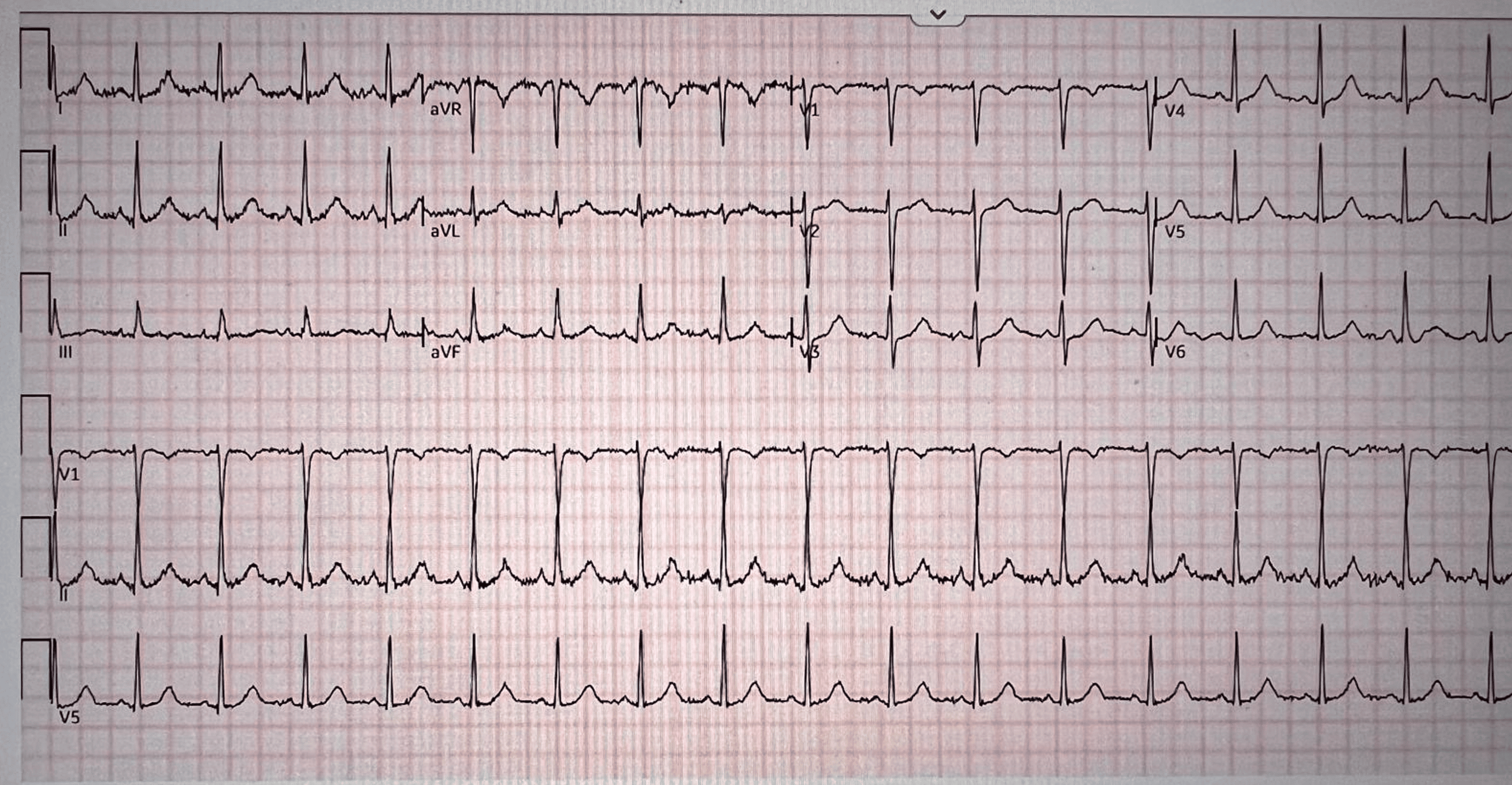
Lab also revealed troponemia of 0.064 ng/mL, which was followed up by electrocardiography (EKG), which showed sinus tachycardia and was otherwise benign (Figure 1).
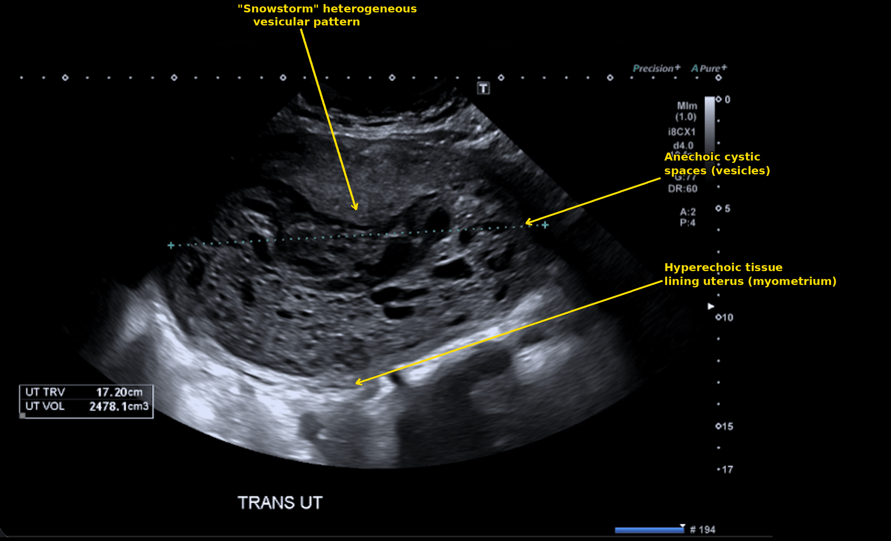
A transvaginal ultrasound revealed the uterus measuring up to 22.6×12.2×17.2 cm. It appeared complex with a mildly thickened endometrium, which was seen extending into the myometrium. It raised concerns for retained products of conception, possibly myometrial invasion, which is seen in GTD. The patient was given three units of packed red blood cells (PRBCs), given the hemorrhage shock secondary to GTD. She was also started on vancomycin 1 gm in 500 mL of sodium chloride (NaCl), metronidazole/sodium chloride (Flagyl in normal saline) 500 mg in 100 mL, and aztreonam 1 mg in 50 mL of dextrose. The gynecology team performed the suction dilatation and curettage (D&C) procedure the same day as admission. The transvaginal ultrasound image is presented in Figure 2.
During the preoperative anesthesia evaluation, the patient was noted to be a high surgical risk candidate with the American Society of Anesthesiologists Physical Status Classification System (ASA classification) of 4E, which is labeled as an emergency procedure for a patient with severe systemic disease that is a constant threat to life [4]. She was planned to undergo general anesthesia via rapid sequence intubation (RSI) due to heightened surgical risk. The anesthesiologist classified the Mallampati score as II, which is labeled as soft palate and uvula visible while pillars are obscured [5]. The patient’s thyromental distance was noted to be three finger breadths. During the intraoperative phase, the patient received three units of PRBCs, two units of fresh frozen plasma (FFP), and one unit of platelets with 400 cc of blood loss. She was transferred to the intensive care unit (ICU) for heightened management of hemorrhage shock and postoperative care. The postoperative complete blood count (CBC) showed a 10.4 g/dL status post five units of blood in total. The patient continued metronidazole, vancomycin, and aztreonam for treating possible sepsis since the white blood cell (WBC) count was elevated to 15.3×10³/µL. She was seen to have minimal urine output via a Foley catheter, possibly secondary to severe hemorrhage. Concerning postoperative labs included low calcium of 7.2 mg/dL, which was likely due to massive transfusion, and low activated partial thromboplastin time (APTT) of 25.6 seconds. β-hCG was seen to be significantly decreasing postoperatively and dropped to 700,000 mIU/mL on post-op day 0 (POD 0). The patient was hemodynamically stable and discharged on POD 3 with weekly outpatient follow-up to trend decreasing β-hCG.
The differential diagnosis for heavy vaginal bleeding in this patient included retained products of conception following her recent missed abortion, endometritis, uterine atony, and GTD. Endometritis was considered less likely given the absence of fever, uterine tenderness, or purulent discharge on presentation, despite mild neutrophilia, which was thought to reflect a physiologic stress response to acute hemorrhage rather than active infection. Simple retained products of conception were considered less likely given the degree of uterine enlargement (22.6×12.2×17.2 cm) and the ultrasound finding of a complex, thickened endometrial stripe extending into the myometrium, a pattern more consistent with trophoblastic tissue invasion than with residual tissue from a missed abortion alone. GTD was ultimately favored based on the combination of these sonographic findings with a markedly elevated β-hCG of 1,889,800 mIU/mL, a level far exceeding what would be expected from retained products of conception or a normal early pregnancy, and prompted urgent gynecology evaluation and D&C.
Discussion
Our case report highlights a complex case of an obstetric and gynecologic emergency of a complete molar pregnancy. Hemorrhage shock due to massive blood loss warranted increased preoperative management of thyroid function, intraoperative airway management, and prevention of postoperative complications of thyroid storm and DIC.
The patient’s markedly elevated β-hCG (1,889,800 mIU/mL) also placed her at substantial risk for thyroid storm, a rare but potentially fatal perioperative complication. Several case reports document thyroid storm in GTD patients undergoing evacuation; Kofinas et al. reported a similarly severe case of thyroid storm-induced multiorgan failure in a 45-year-old patient with GTD and a β-hCG of 1,488,021 mIU/mL, closely comparable to this patient’s presentation [6]. Intraoperative vigilance is critical, and anesthesia providers should maintain a high index of suspicion for thyroid storm in patients with persistent tachycardia, hypertension, or hyperthermia unresponsive to standard interventions. Interestingly, our patient’s thyroid function tests showed an elevated TSH (5.92 mIU/L) with a normal free T4 (0.93 ng/dL), a pattern inconsistent with the biochemical hyperthyroidism classically described in hCG-mediated thyroid stimulation. Levothyroxine, part of her home regimen for known hypothyroidism, was held as part of the routine perioperative management of home medications during the acute surgical emergency and was resumed once β-hCG levels began to trend downward following D&C. This underscores that thyroid storm risk in GTD may not always be reflected on standard thyroid function testing and that clinical vigilance for signs of thyrotoxicosis (tachycardia, hyperthermia, hypertension) remains essential regardless of TSH/free T4 values.
Several aspects of anesthetic management in this case merit discussion in the context of available alternatives. The literature reflects a range of anesthetic approaches to molar pregnancy evacuation, and the optimal technique remains debated. A retrospective analysis of 110 molar pregnancy patients found that general anesthesia was more likely to be selected in patients with larger uterine size, hyperthyroidism, and higher serum β-hCG levels, though intraoperative complications such as uterine atony, massive hemorrhage, and pulmonary edema were observed only in the general anesthesia group [7]. Spinal and combined spinal-epidural anesthesia have also been described in this population and may offer a theoretical advantage in patients with biochemical hyperthyroidism by avoiding the sympathetic stimulation associated with laryngoscopy and intubation, provided hemodynamic stability is maintained [8]. Total intravenous anesthesia has similarly been reported as an alternative to volatile agent-based general anesthesia for the evacuation of a hydatidiform mole in a thyrotoxic patient [9]. In our patient, active hemorrhage, hemodynamic instability, and the need for a definitively secured airway favored general anesthesia over neuraxial techniques, consistent with the pattern of larger uterine size, elevated β-hCG, and greater comorbidity burden favoring general anesthesia reported in the literature.
We used RSI during general intubation for this case, and it was indicated for several reasons. First, all recently pregnant patients (within 48 hours of delivery or pregnancy loss) are considered to have increased aspiration risk due to delayed gastric emptying, decreased lower esophageal sphincter tone, and increased intra-abdominal pressure from the gravid uterus [10]. Although this patient presented two months after her previous failed pregnancy, the presence of retained products of conception and ongoing pregnancy-related hormonal changes may have maintained some degree of aspiration risk. Additionally, the urgent nature of the surgical procedure precluded prolonged preoperative fasting or gastric emptying. Second, the patient’s hemodynamic instability from massive hemorrhage and severe anemia necessitated rapid control of the airway to ensure adequate oxygenation and ventilation. RSI allows for the fastest possible transition from spontaneous ventilation to controlled mechanical ventilation with a secured airway. Furthermore, two large-bore intravenous access lines, type and crossmatch, and availability of a massive transfusion protocol were ensured preoperatively.
This report has several limitations inherent to single-case reporting. As a single case, the findings and management approach described here cannot be generalized to all patients with GTD-associated hemorrhage or thyrotoxicosis, and outcomes may vary based on institutional resources, patient comorbidities, and disease severity. The retrospective nature of this report also limits the ability to establish causal relationships between specific interventions and outcomes; the favorable outcome in this case cannot be definitively attributed to any single management decision. Additionally, the full trajectory of thyroid function throughout the perioperative period, and its relationship to the declining β-hCG level, was not comprehensively characterized in this report. Larger case series or prospective studies would be needed to better define optimal perioperative thresholds and management protocols for this rare but high-acuity patient population.
This case underscores several key clinical lessons for anesthesiologists managing patients with GTD. First, extreme β-hCG elevation should prompt a high index of suspicion for both hemorrhagic coagulopathy and thyroid storm, even in the absence of confirmatory biochemical hyperthyroidism, as thyroid function testing may not reliably capture this risk. Second, anesthetic technique selection should be individualized, weighing the airway control and hemorrhage-response advantages of general anesthesia against the potential sympathetic stimulation risks in thyrotoxic-prone patients. Third, close perioperative laboratory and clinical monitoring, along with preparedness for massive transfusion, are essential even when overt DIC or thyroid storm do not ultimately develop. A coordinated, multidisciplinary approach involving obstetrics, gynecology, and anesthesiology is critical to optimizing outcomes in this rare but potentially life-threatening presentation.
Conclusions
This case illustrates the complex anesthetic challenges posed by GTD with massive hemorrhage. The extremely elevated β-hCG level created a risk for thyroid storm, while massive transfusion predisposed to DIC. RSI was appropriately indicated given the aspiration risk and hemodynamic instability. Successful management requires preoperative risk stratification including thyroid function testing, meticulous intraoperative monitoring for complications, aggressive hemorrhage control with balanced transfusion protocols, and vigilance for thyroid storm manifestations. A multidisciplinary approach involving anesthesia, obstetrics, and critical care is essential for optimal outcomes in these high-risk patients. As these observations are drawn from a single case, they are intended to highlight important clinical considerations for anesthesiologists managing similar presentations rather than to establish the superiority of any particular management strategy. Thus, larger case series are needed to validate these approaches more broadly.
References
- Horowitz NS, Eskander RN, Adelman MR, Burke W: Epidemiology, diagnosis, and treatment of gestational trophoblastic disease: a Society of Gynecologic Oncology evidenced-based review and recommendation. Gynecol Oncol. 2021, 163:605-13. 10.1016/j.ygyno.2021.10.003
- Cunningham FG, Nelson DB: Disseminated intravascular coagulation syndromes in obstetrics. Obstet Gynecol. 2015, 126:999-1011. 10.1097/AOG.0000000000001110
- ACOG practice bulletin no. 209: obstetric analgesia and anesthesia. Obstet Gynecol. 2019, 133:e208-25. 10.1097/AOG.0000000000003132
- American Society of Anesthesiologists statement on ASA physical status classification system. Anesthesiol Open. 2026, 1:e0002. 10.1097/ao9.0000000000000002
- Jackson JS, Rondeau B: Mallampati score. StatPearls [Internet]. StatPearls Publishing, Treasure Island (FL); 2026.
- Kofinas JD, Kruczek A, Sample J, Eglinton GS: Thyroid storm-induced multi-organ failure in the setting of gestational trophoblastic disease. J Emerg Med. 2015, 48:35-8. 10.1016/j.jemermed.2014.09.012
- Triyasunant N, Nivatpumin P, Luangpirom N, Wongwananuruk T: Factors influencing decision-making for the anesthetic technique in molar pregnancy patients for suction curettage: a retrospective study. Indian J Clin Anaesth. 2024, 11:25-31. 10.18231/j.ijca.2024.006
- Swaminathan S, James RA, Chandran R, Joshi R: Anaesthetic implications of severe hyperthyroidism secondary to molar pregnancy: a case report and review of literature. Anesth Essays Res. 2017, 11:1115-7. 10.4103/aer.AER_38_17
- Erturk E, Bostan H, Geze S, Saracoglu S, Erciyes N, Eroglu A: Total intravenous anesthesia for evacuation of a hydatidiform mole and termination of pregnancy in a patient with thyrotoxicosis. Int J Obstet Anesth. 2007, 16:363-6. 10.1016/j.ijoa.2006.12.004
- Sorbello M, Paternò DS, Zdravkovic I, La Via L: Pharmacological approach to rapid sequence induction/intubation: a contemporary perspective. Curr Opin Anaesthesiol. 2025, 38:369-74. 10.1097/ACO.0000000000001528