

Authors Saring N, Ado P, Sanyal R
Cureus 18(10): e118357. doi:10.7759/cureus.118357
Abstract
Introduction: Noninvasive monitoring methods are increasingly being explored to avoid complications associated with invasive techniques in perioperative or critical care settings for optimal hemodynamic management. The perfusion index (PI) derived from pulse oximetry reflects peripheral perfusion, while the inferior vena cava (IVC)-collapsibility index (CI) measured by ultrasonography is a useful indicator of intravascular volume status. The present study aimed to evaluate the correlation between PI and IVC-CI and to assess their relationship with mean arterial pressure (MAP).
Methods: This prospective analytic study included adult preoperative patients with American Society of Anesthesiologists (ASA) physical status I and II who fulfilled the inclusion criteria. Perfusion index was recorded using pulse oximetry, and IVC diameters were measured using bedside ultrasonography to calculate IVC-CI. Mean arterial pressure was estimated using noninvasive blood pressure measurement. Spearman’s correlation coefficient was used for the determination of the correlation between PI and IVC-CI, MAP and PI, and MAP and IVC-CI.
Results: A total of 119 adult patients aged 33.06 ± 12 (mean ± SD) years, belonging to ASA physical status I and II, and posted for elective surgical procedures were included in this study. The baseline MAP was 92.98 ± 11.32 mmHg. There were no participants with PI < 0.03, and the baseline PI was 2.63 ± 2.11 (mean ± SD). The preoperative mean ± SD maximum (IVCmax) and minimum (IVCmin) IVC diameter measurements were 1.58 ± 1.36 and 1.03 ± 1.41 cm, respectively. The correlation coefficient (r) was -0.012 between PI and IVC-CI (p = 0.893), -0.070 between PI and MAP (p = 0.447), and 0.054 between IVC-CI and MAP (p = 0.561).
Conclusions: The present study found no significant correlation between PI and IVC-CI. A positive correlation was observed between IVC-CI and MAP, in contrast to the inverse relationship observed between PI and MAP, but these findings were not statistically significant.
Introduction
Optimal assessment of intravascular fluid status is a cornerstone of safe anesthetic and perioperative management. Both hypovolemia and fluid overload are associated with adverse outcomes considering the fasting status of patients along with the administration of anesthetic agents, positive pressure ventilation, and presence of comorbidities. Hypovolemia may lead to hypotension and impaired organ perfusion, while hypervolemia may lead to edema, oxygen desaturation, and organ dysfunction. Despite its clinical importance, an accurate assessment of intravascular volume remains challenging. The conventional clinical parameters such as heart rate, blood pressure, urine output, and central venous pressure have limited sensitivity and specificity and often fail to reflect true volume status in most cases [1,2].
Ultrasonography is a noninvasive, dynamic, economic, and integral part of modern anesthetic practice, with applications ranging from vascular access, regional anesthesia, gastric content assessment, airway assessment, hemodynamic evaluation, and focused thoracic echocardiography [3-5]. Respiratory variation in the inferior vena cava (IVC) diameter reflects significant changes in venous return and right atrial pressure (RAP) due to its thin-walled and highly compliant vessel, particularly in the spontaneously breathing patients [6]. The IVC collapsibility index (IVC-CI) calculated from inspiratory (minimum) and expiratory (maximum) diameters, has been incorporated into several point-of-care ultrasound (POCUS) protocols and is increasingly used by anesthesiologists and critical care providers [7,8].
Photoplethysmography (PPG) is a simple, inexpensive, continuous, and noninvasive technique that is universally available in operating rooms as part of the minimum standard of monitoring guideline by the American Society of Anesthesiologists (ASA) [9]. It forms the basis of pulse oximetry. Perfusion index (PI) values are the ratio of pulsatile blood flow to non-pulsatile blood flow in the peripheral tissues, which vary widely among healthy individuals, typically ranging from 0.3 to 10.0, with a reported median value of 1.4. Since PI reflects changes in the peripheral vascular tone and the stroke volume, it may be influenced by the intravascular volume status and thus may have a potential role as a noninvasive surrogate marker for the fluid status assessment [10-12].
Although both PI and IVC-CI have independently shown promise as tools for intravascular volume assessment, limited data exist correlating these two noninvasive parameters, especially in the perioperative elective surgical population. Therefore, this prospective analytical study was aimed at evaluating the correlation between PI and IVC-CI in adult patients aged 18 to 60 years undergoing elective surgery and also determining the correlation between preoperative MAP and PI and preoperative MAP and IVC-CI.
Materials & Methods
Study design and setting
This prospective analytic study was conducted in the Department of Anesthesiology and Critical Care, Tomo Riba Institute of Health and Medical Sciences (TRIHMS) (Naharlgun, AP, IND) after obtaining approval from the Institutional Ethics Committee (approval no. TRIHMS/IEC/05/2023/676 dated 02/04/2024). The study was carried out over a period of one and a half years, from April 2024 to October 2025.
Study population and sampling
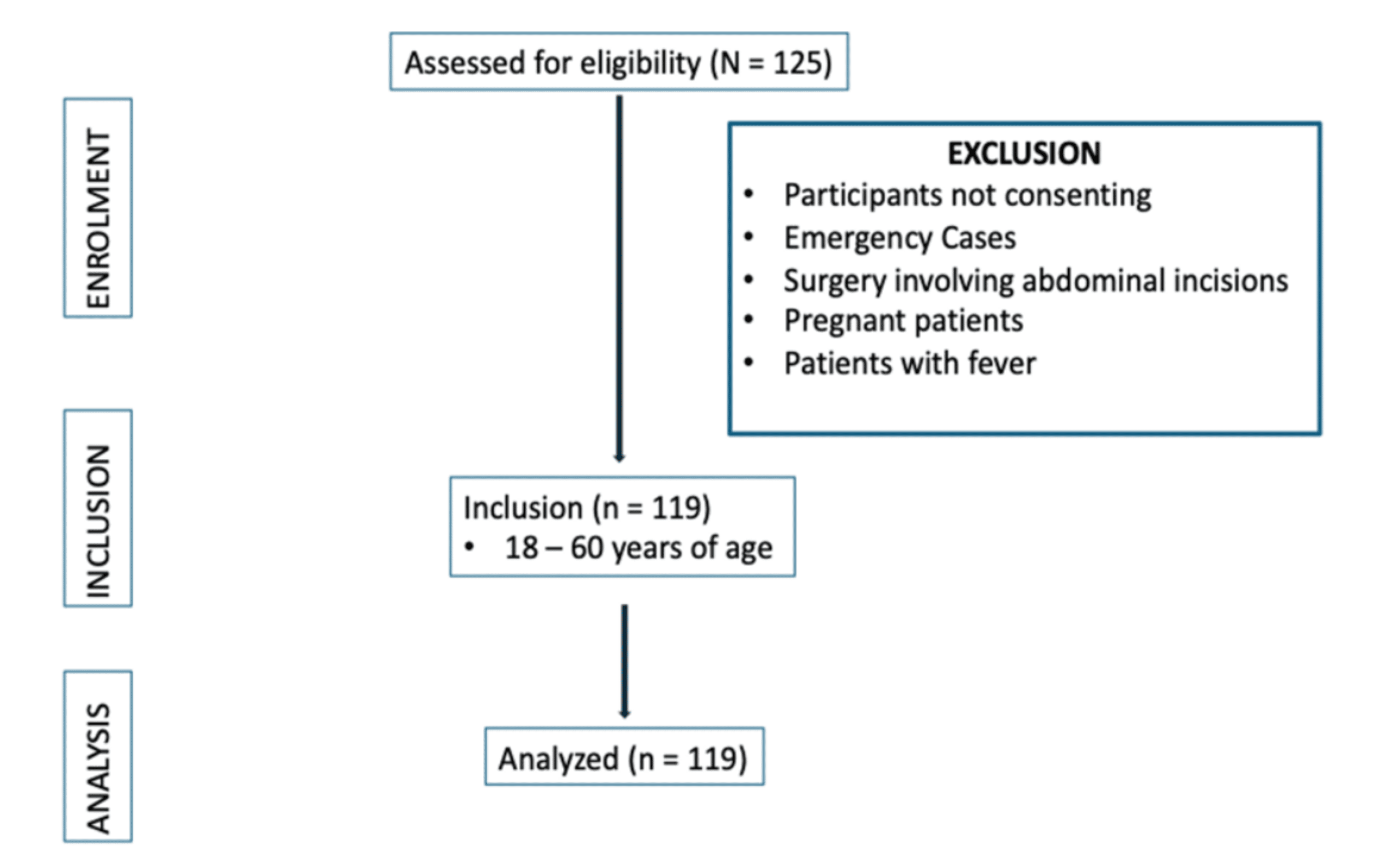
A total of 119 patients scheduled for elective surgical procedures were analyzed in the study. Consenting adult patients of 18 to 60 years of age with ASA physical status I and II posted for elective surgery under general anesthesia were included in the study. Patients posted for an emergency surgery, pregnant, or presenting with signs of acute infection were excluded from the study. The consecutive sampling technique was used. Sample size was calculated to be 113 with a correlation coefficient of 0.30 (derived from a previous study [13]), with a CI of 95%, and a power of 90%. A total of 119 patients were enrolled in the study (Figure 1).
Study procedure
The patients posted for routine surgery were subjected to preanesthetic evaluation, and those fulfilling the inclusion criteria were enrolled in the study after written informed consent. The participants abstained from solid food for at least eight hours and clear fluids for at least two hours before surgery as per institutional protocol. On the day of the surgery, before initiation of any parenteral therapy, the standard mandatory ASA monitoring was applied, and the parameters, including pulse rate, noninvasive blood pressure (NIBP), temperature, and peripheral oxygen saturation (SpO2), were recorded in the supine position. The study proceeded if participants had normal temperature, pulse rate, MAP, and SpO2. The PI and IVC-CI were measured by an anesthesiologist.
The PI was measured using the plethysmography probe of the Philips Goldway GS20, which is available at our institution free of cost (Philips Goldway (Shenzhen) Industrial Inc., Shenzhen, GD, CHN). The probe was attached to the index finger in the supine position after it was screened for ambient light. A single PI reading was obtained after maintaining a stable plethysmographic waveform for at least one minute during quiet normal breathing of ambient air.
The IVC measurements were performed using an ACUSON P500 (Siemens Healthineers, Erlangen, DEU) ultrasound machine (available at the institution free of cost) with a curvilinear probe (frequency bandwidth: 1.4-5.0 MHz). With the patient in the supine position, the probe was placed in the sub-xiphoid region to obtain a long-axis view of the IVC. A reading of the maximum (expiratory) and minimum (inspiratory) IVC diameters was measured using M-mode ultrasonography approximately 1 cm distal to the hepatic vein inlet over a single respiratory cycle of quite normal breathing of ambient air. The IVC-CI was calculated using the formula IVCCI=IVCmax−IVCminIVCmax. The goal for IVC-CI was taken from 20% to 50% [7], respecting potential biases in interpretation and overriding clinical considerations. After measurement of the parameters mentioned above, the participants were subjected to routine procedures, and the study was deemed complete.
Statistical analysis
The data were analyzed via SPSS Statistics version 25.0 (IBM Corp., Armonk, NY, USA). The parameters were recorded as mean and SD for continuous variables, and frequency and percentage for categorical variables. The correlation between PI and IVC-CI, PI and MAP, and IVC-CI and MAP was assessed using Spearman’s rho correlation coefficient.
Results
A total of 119 adult patients aged 18 to 60 years with ASA physical status I and II undergoing elective surgical procedures were enrolled in the study. The mean ± SD of age of subjects was 33.06 ± 12 (range: 18 to 60) years, with the majority aged between 21 and 30 years (45, 37.82%); 54 (45.38%) of the participants were female, while 65 (54.62%) were males (Table 1). The mean ± SD height was 157.07 ± 8.02 cm, while the mean ± SD weight was 58.38 ± 9.23 kg, and the mean ± SD BMI was 23.63 ± 3.07 kg/m²; 68 (57.14%) of the participants had a normal BMI (range of 18.5 to 24.99 kg/m²), while seven (5.88%) were underweight (<18.5 kg/m²), 43 (36.13%) were overweight (25 to 29.99 kg/m²), and one (0.84%) was obese (≥30 kg/m²).
The participants had a normal temperature, with an SpO2 of >95%, and a mean pulse rate of 82 ± 8.13 beats per minute. The preoperative systolic blood pressure (SBP) was 122.91 ± 10.5 (median: 123) mmHg, diastolic blood pressure (DBP) 77.43 ± 11.18 (median: 76) mmHg, and MAP 92.98 ± 11.32 (median: 92) mmHg (Table 2).
The preoperative PI was within the normal range in all participants. The mean ± SD PI was 2.63 ± 2.11, with a median of 1.7 (0.95-3.8). The preoperative IVCmax had a mean ± SD of 1.58 ± 1.36 cm (median: 1.55 (1.5-1.67) cm) and IVCmin had a mean ± SD of 1.03 ± 1.41 cm (median: 1.01 (0.95-1.13) cm). The preoperative IVC-CI was normal in 117 (98.32%) participants and deranged in two (1.68%) participants. The mean ± SD IVC-CI was 34.76 ± 7.03, with a median (25th-75th percentile) of 34.46 (30.17-40.125).
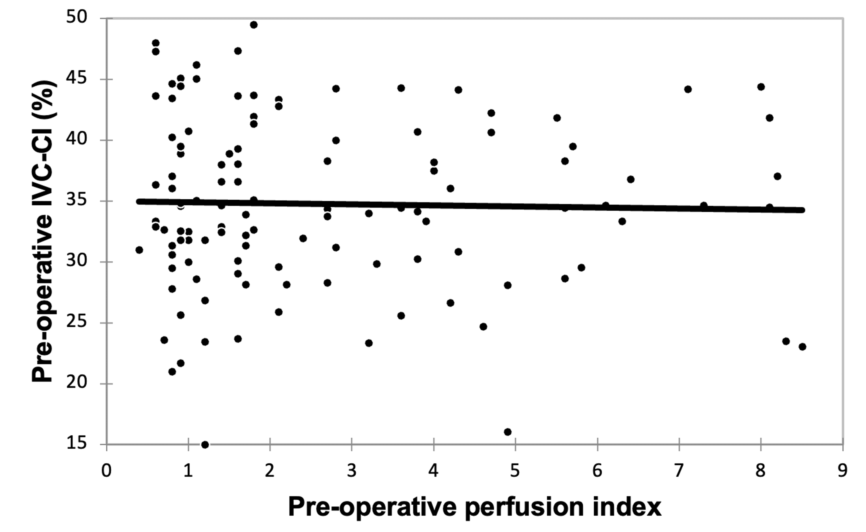
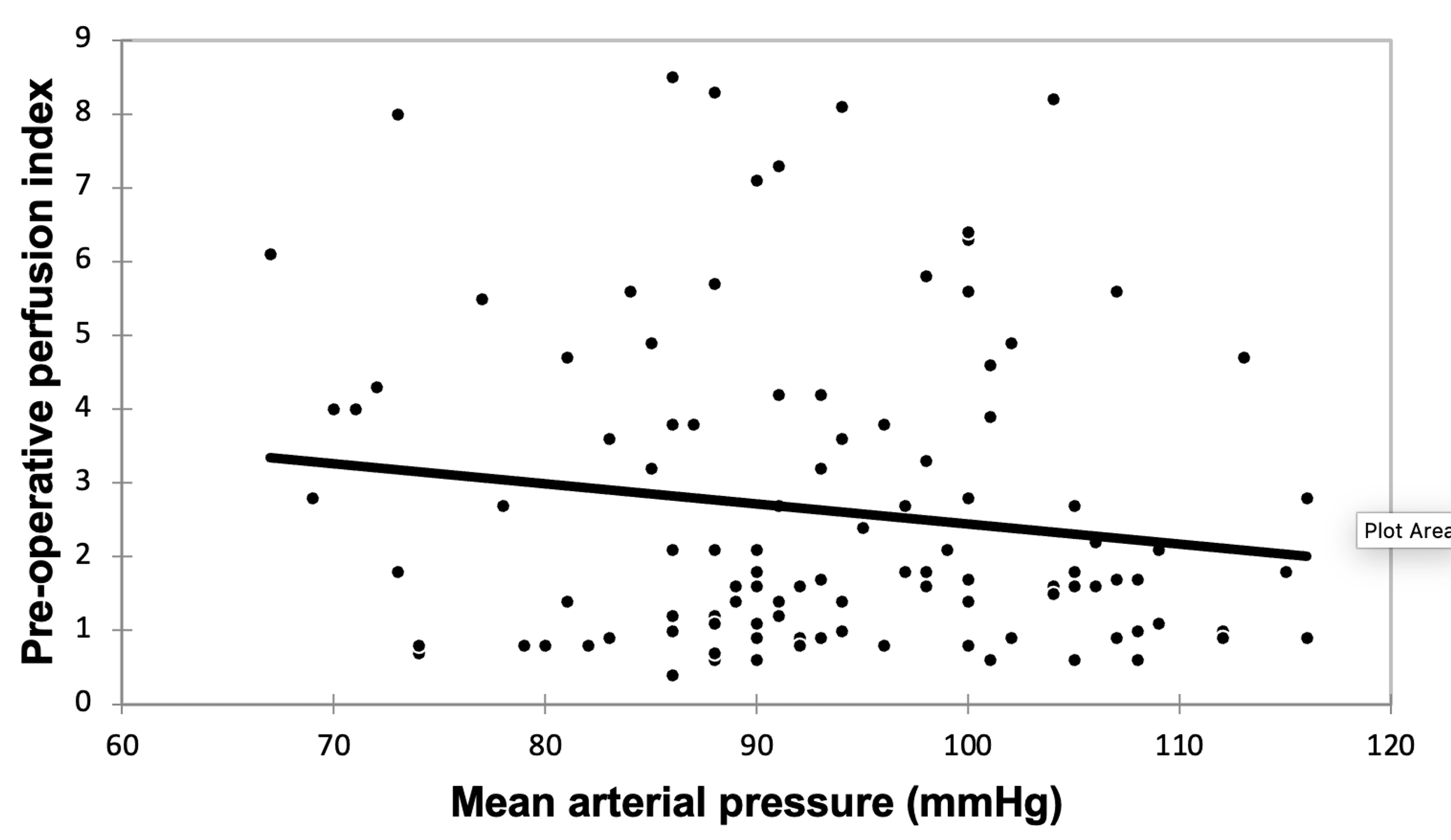
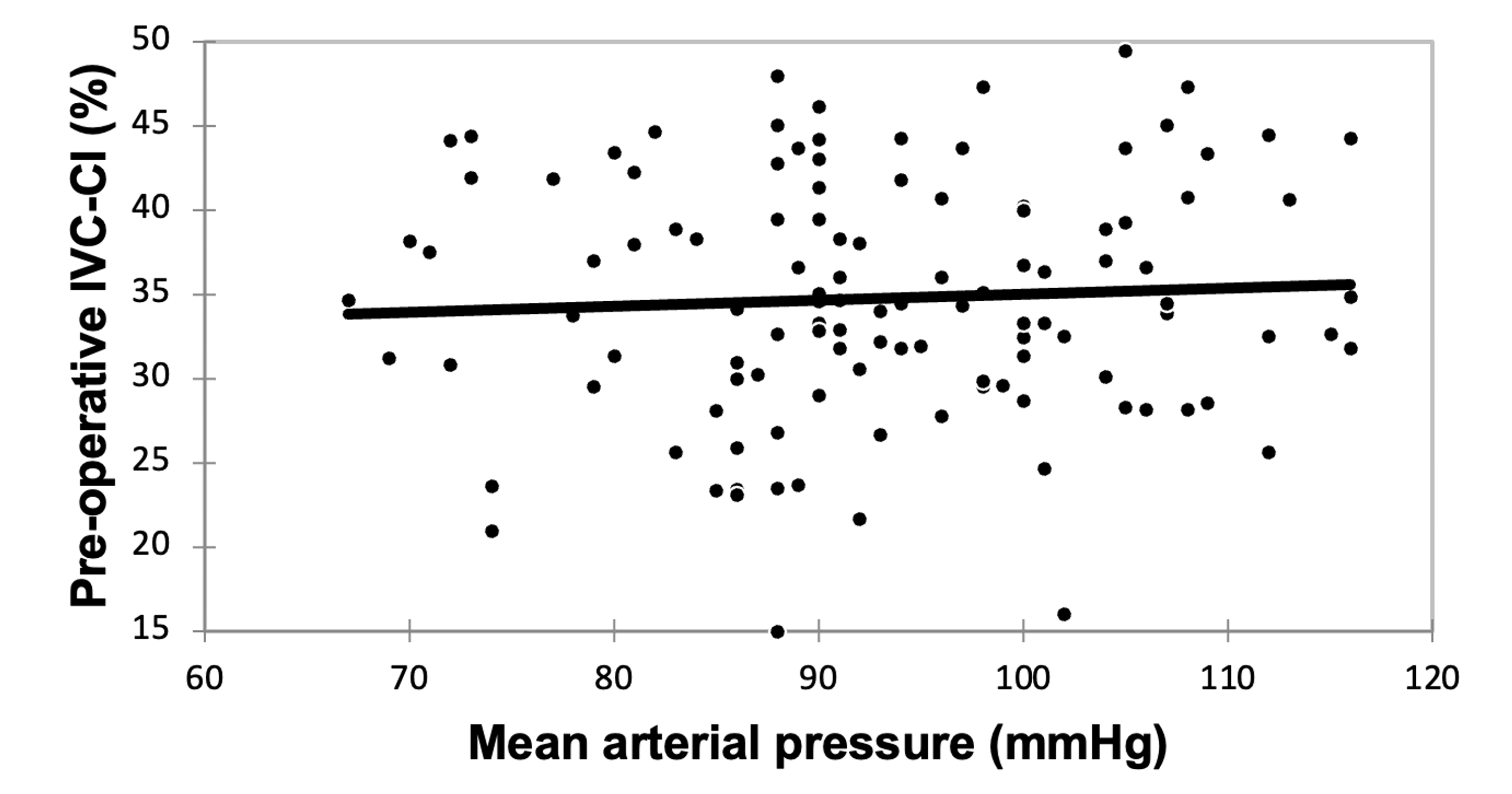
The Spearman rank correlation coefficient between PI and IVC-CI was found to be -0.012 with a p-value of 0.893 (Table 3 and Figure 2). A very weak negative correlation was observed between preoperative PI and preoperative MAP (r = −0.070, p-value = 0.447) (Figure 3), while a very weak positive correlation was observed between preoperative IVC-CI (%) and preoperative MAP (mmHg), with r = 0.054 and p-value = 0.561 (Figure 4).
Discussion
Perfusion index
In the present study, the preoperative PI was within the normal range in all participants, with a mean of 2.63 ± 2.11. The wide range observed (0.4-8.5) reflects the known variability of PI [10], which is influenced by multiple factors, including peripheral vasomotor tone, ambient temperature, anxiety, and sympathetic activity [14]. Despite this variability, all values were considered clinically acceptable, suggesting adequate peripheral perfusion in the study cohort.
IVC measurements and CI
The mean IVCmax and IVCmin values were 1.58 ± 1.36 cm and 1.03 ± 1.41 cm, respectively, resulting in a mean IVC-CI of 34.76 ± 7.03%. Nearly all participants (98.32%) demonstrated a normal IVC-CI, indicating euvolemic status in the majority of cases, as the participants enrolled in the study were posted for elective surgery and ASA physical status I and II, who are adequately optimized preoperatively.
Correlation between PI and IVC-CI
A very weak negative correlation was observed between preoperative PI and IVC-CI (r = -0.012, p = 0.893), which was not statistically significant. This finding was not in congruence with the study findings of Arac et al. [13], where the authors found a significant inverse relationship between PI and IVC-CI in participants in a fasting state. Perfusion index is considered an indirect indicator of peripheral tissue perfusion and microcirculatory blood flow, as it is obtained by the change in the blood volume of peripheral vessels in each pulse, thereby influenced by physiological and environmental factors such as cardiac output, blood volume, perfusion pressure, and systemic vascular tone. Factors like sympathetic nervous system activity, peripheral vasomotor disorders, temperature, anxiety, and pain can also lead to significant changes in peripheral vascular tone [14,15]. On the other hand, the IVC diameter measurement and the CI are commonly used as a noninvasive indicator of intravascular volume status and right atrial pressure. Ultrasound evaluation of IVC-CI is easy to obtain, non-invasive, cheap, and easy to calculate [16].
Perfusion index and IVC-CI represent distinct components of the cardiovascular system. The changes in the IVC-CI reflect central venous capacitance and preload status through dynamic changes in its anatomy during the respiratory cycle. Whereas the PI of plethysmography reflects peripheral microcirculatory perfusion, which is based on the pulse changes of regional area blood flow or volume [17]. Because these parameters originate from different physiological domains, changes in one parameter may not necessarily correspond to changes in the other. The observation that there is no correlation between the PI and IVCCI in the current study is not congruent with the findings of Guduk et al.’s study [15], as well as the study by Kesavankutty et al. [18], as one found a negative and the other found a positive statistical (r = 525, p = 0.003) correlation between PI and ICV-CI.
The absence of correlation observed in the study may also be explained by the clinical characteristics, as both PI and IVC-CI are likely to remain within normal physiological ranges with limited variability. Another possible explanation is the influence of sympathetic tone on peripheral perfusion. Anxiety, mild stress, or environmental factors in the preoperative setting may lead to peripheral vasoconstriction, thereby reducing PI values even when intravascular volume is adequate [14]. Conversely, mild peripheral vasodilation may increase PI without altering central venous volume. Such local vascular influences may weaken the relationship between PI and IVC-CI. Therefore, the lack of significant correlation between PI and IVC-CI in the present study may likely reflect the distinct physiological processes represented by these indices, combined with the relatively homogeneous and hemodynamically stable nature of the study population. Rather than being interchangeable, these parameters should be viewed as complementary tools for evaluating different aspects of circulatory function.
Correlation between MAP and PI
In the present study, a very weak negative correlation is observed between MAP and PI, but this finding is not statistically significant (r = -0.070, p = 0.447). Unlike systemic blood pressure, which reflects the overall balance between cardiac output and systemic vascular resistance, PI primarily reflects microcirculatory blood flow at the peripheral tissue level, which may be physiologically influenced by the sympathetic tone on peripheral circulation. An increase in sympathetic activity can lead to vasoconstriction and thereby lower PI values, even when the systemic blood pressure remains within normal limits. Consequently, the changes in PI may occur independently of the systemic arterial pressure [19-21]. The MAP is tightly regulated by compensatory cardiovascular mechanisms, renal and autonomic modulation. In relatively healthy individuals such as the ASA I and II population, these compensatory mechanisms may maintain stable BP despite minor fluctuations in peripheral perfusion. Therefore, PI may vary due to local vascular factors without producing measurable changes in MAP [22]. The absence of significant correlation in this study suggests that PI should not be interpreted as a surrogate marker of systemic arterial pressure. Rather, it provides complementary information regarding the peripheral perfusion and the microvascular circulation, which may not always parallel systemic hemodynamic parameters.
Correlation between MAP and IVC-CI
The study also demonstrated a very weak positive correlation between MAP and IVC-CI (r = 0.054, p = 0.561), which was not statistically significant. In elective surgical patients who are adequately hydrated and clinically stable, compensatory mechanisms may often maintain a normal arterial pressure even when there are mild variations in the intravascular volume [22]. Additionally, the predominance of euvolemic patients with normal IVC-CI values may likely limit the variability required to detect a meaningful correlation with MAP.
The PI and IVC-CI provide valuable information regarding circulatory physiology, but they do not necessarily correlate with each other and with systemic arterial pressure in hemodynamically stable patients. Thereby, the findings of this study suggest that PI cannot be used as a standalone surrogate for intravascular volume assessment in preoperative elective surgical patients. Similarly, while IVC-CI remains a useful non-invasive tool for assessing volume status, it should be used in conjunction with clinical assessment and other hemodynamic parameters. Both indices provide complementary information. The present study therefore adds to the growing body of evidence indicating that PI and IVC-CI represent distinct physiological domains of hemodynamic monitoring, and their combined interpretation may offer a more comprehensive assessment of intravascular volume and tissue perfusion.
Limitations
The study involves subjects with ASA physical status I and II, with predominant euvolemic status in a single-center study that limits generalizability, especially in patients with hypovolemia as well as fluid responsiveness. The single preoperative measurement of both PI and IVC-CI did not evaluate the dynamic changes in these indices following fluid administration or physiological stress, which may have provided better insight into their relationship. Although measurements were taken under standardized conditions, the PI could have been influenced by confounding factors such as ambient temperature, patient anxiety, sympathetic tone, and peripheral vasoconstriction.
Conclusions
The present study found no significant correlation between PI and IVC-CI in predominantly euvolemic preoperative patients. These findings suggest that PI alone may not reliably reflect intravascular volume status when compared with IVC-CI in stable preoperative patients. The use of multimodal assessment cannot be overemphasized. The PI and IVC-CI should be used as complementary tools.
References
- Singh S, Kuschner WG, Lighthall G: Perioperative intravascular fluid assessment and monitoring: a narrative review of established and emerging techniques. Anesthesiol Res Pract. 2011, 2011:231493. 10.1155/2011/231493
- Marik PE: Fluid responsiveness and the six guiding principles of fluid resuscitation. Crti Care Med. 2016, 44:1920-1922. 10.1097/CCM.0000000000001483
- Gupta P, Gupta K, Dwivedi AN, et al.: Potential role of ultrasound in anesthesia and intensive care. Anesth Essays Res. 2011, 5:11-19. 10.4103/0259-1162.84172
- Terkawi AS, Karakitsos D, Elbarbary M, Blaivas M, Durieux ME: Ultrasound for the anesthesiologists: present and future. Sci World J. 2013, 2013:683685. 10.1155/2013/683685
- Di Nicolò P, Tavazzi G, Nannoni L, Corradi F: Inferior vena cava ultrasonography for volume status evaluation: an intriguing promise never fulfilled. J Clin Med. 2023, 12:10.3390/jcm12062217
- Natori H, Tamaki S, Kira S: Ultrasonographic evaluation of ventilatory effect on inferior vena caval configuration. Am Rev Respir Dis. 1979, 120:421-427. 10.1164/arrd.1979.120.2.421
- Kaptein MJ, Kaptein EM: Inferior vena cava collapsibility index: clinical validation and application for assessment of relative intravascular volume. Adv Chronic Kidney Dis. 2021, 28:218-226. 10.1053/j.ackd.2021.02.003
- Beigel R, Cercek B, Luo H, Siegel RJ: Noninvasive evaluation of right atrial pressure. J Am Soc Echocardiogr. 2013, 26:1033-1042. 10.1016/j.echo.2013.06.004
- Elsevier: Miller’s Anesthesia, 2-Volume Set. Gropper MA, Cohen NH, Eriksson LI, et al. (ed): Elsevier, 2024. https://shop.elsevier.com/books/millers-anesthesia-2-volume-set/gropper/978-0-323-93592-0.
- Lima AP, Beelen P, Bakker J: Use of a peripheral perfusion index derived from the pulse oximetry signal as a noninvasive indicator of perfusion. Crit Care Med. 2002, 30:1210-1213. 10.1097/00003246-200206000-00006
- Elgendi M: Optimal signal quality index for photoplethysmogram signals. Bioengineering. 2016, 3:4. 10.3390/bioengineering3040021
- Coutrot M, Dudoignon E, Joachim J, Gayat E, Vallée F, Dépret F: Perfusion index: Physical principles, physiological meanings and clinical implications in anaesthesia and critical care. Anaesth Crit Care Pain Med. 2021, 40:100964. 10.1016/j.accpm.2021.100964
- Arac S, Ozel M, Zengin S, Kanbur I, Tahir Gokdemir M: Use of vena cava inferior collapsibility index and perfusion index in volume status monitoring during intermittent fasting in the ramadan. Ann Med Res. 2021, 5:0957-0962.
- Sun X, He H, Xu M, Long Y: Peripheral perfusion index of pulse oximetry in adult patients: a narrative review. Eur J Med Res. 2024, 29:457. 10.1186/s40001-024-02048-3
- Güdük NK, Sahin T: Follow-up of perfusion index and inferior vena cava collapsibility index in fluid therapy in prerenal acute renal failure. Heliyon. 2023, 9:e18715. 10.1016/j.heliyon.2023.e18715
- Park JC, Yun SH: Analysis of patterns of pulse oximeter Perfusion Index according to postural change during gynecological surgery. J Cell Mol Anesth. 2025, 10:162363. 10.5812/jcma-162363
- Pyakurel K, Rajbanshi LK, Khanal K, et al.: Correlation study of inferior venacava collapsibility index and perfusion index as predictors of fluid responsiveness in critically ill patients. Birat J Health Sci. 2023, 8:2167-2173. 10.62065/bjhs478
- Kesavankutty MP, Panda C, Karim HMR, Singha S, Agrawal S: The diagnostic accuracy of preoperative perfusion index as a predictor of post-spinal anesthesia hypotension in parturients undergoing cesarean delivery: a prospective non-blinded observational study. Saudi J Anaesth. 2024, 18:23-30. 10.4103/sja.sja_378_23
- Caplan M, Durand A, Bortolotti P, et al.: Measurement site of inferior vena cava diameter affects the accuracy with which fluid responsiveness can be predicted in spontaneously breathing patients: a post hoc analysis of two prospective cohorts. Ann Intensive Care. 2020, 10:168. 10.1186/s13613-020-00786-1
- Yang B, Moss J: Evolution of the pulse ox. Chest. 2023, 164:24-26. 10.1016/j.chest.2022.12.042
- Kansiz T, Ayoglu H, Aydin BG, et al.: The efficacy of inferior vena cava diameter, perfusion index and pleth variability index on predicting hypotension in spinal anesthesia. Ann Med Res. 2022, 24:616-622.
- DeMers D, Wachs D: Physiology, Mean Arterial Pressure. StatPearls, Treasure Island; 2026.