

Authors: Goyal M et al
Cureus 18(10): e118126. doi:10.7759/cureus.118126
Abstract
Electrical storm following myocardial infarction (MI) can be difficult to control when antiarrhythmic drugs and cardioversion fail. We describe a 53-year-old man with hypertension and previously undiagnosed diabetes who presented with an inferolateral ST-segment elevation MI caused by total occlusion of the proximal left circumflex artery, which was treated with primary percutaneous coronary intervention and drug-eluting stent placement. He then developed marked leukocytosis with a systemic inflammatory response and acute kidney injury (peak creatinine, 4.03 mg/dL), which resolved with empirical antibiotic therapy and supportive renal care. On day 6, he experienced recurrent syncope associated with sustained monomorphic ventricular tachycardia (VT) that persisted despite intravenous amiodarone and lidocaine, electrolyte correction, and repeated cardioversion, necessitating sedation and mechanical ventilation. Repeat coronary angiography demonstrated a patent stent. VT ceased following an ultrasound-guided left stellate ganglion block (SGB) using 1% lidocaine and dexamethasone, which resulted in Horner syndrome. Antiarrhythmic therapy was continued, and the patient was extubated within 24 hours. Speckle-tracking echocardiography demonstrated severely reduced lateral and anterior strain (global longitudinal strain, -12.0%). A patent stent in the culprit vessel and sustained monomorphic arrhythmia made a scar-related substrate the leading consideration. The close temporal association between the block and cessation of VT suggests that sympathetic blockade may have contributed to arrhythmia control, although the continuation of antiarrhythmic therapy precludes firm causal conclusions. Left SGB may serve as a rapid bridge to definitive treatment in patients with refractory electrical storm.
Introduction
Electrical storm is defined as three or more episodes of sustained ventricular arrhythmia within 24 hours [1]. After myocardial infarction (MI), it carries a high in-hospital mortality, particularly when episodes recur despite antiarrhythmic drugs, beta-blockade, electrolyte correction, and deep sedation [1]. Heightened sympathetic tone both triggers ventricular tachycardia (VT) and makes it harder to suppress [2]. Much of the efferent sympathetic supply to the ventricles passes through the stellate ganglia, so injecting local anesthetic around the left stellate ganglion produces temporary, reversible cardiac sympathetic blockade that can be performed at the bedside. For drug-refractory monomorphic VT storm, current guidelines recommend catheter ablation and suggest that autonomic modulation may be considered when ablation is ineffective or not possible [1]. Percutaneous stellate ganglion block (SGB) has therefore been used mainly as rescue or bridging therapy rather than as a substitute for definitive treatment [2-5]. We report a patient in whom monomorphic VT storm developed four days after primary percutaneous coronary intervention (PCI) for an occluded left circumflex artery (LCx), despite a patent stent, while he was recovering from a systemic inflammatory illness with acute kidney injury (AKI). VT ceased after left SGB with short-acting lidocaine while antiarrhythmic therapy continued. The case illustrates the bedside distinction between stent failure and a scar-related mechanism, and the use of SGB as a bridge when conventional therapy has failed.
Case Presentation
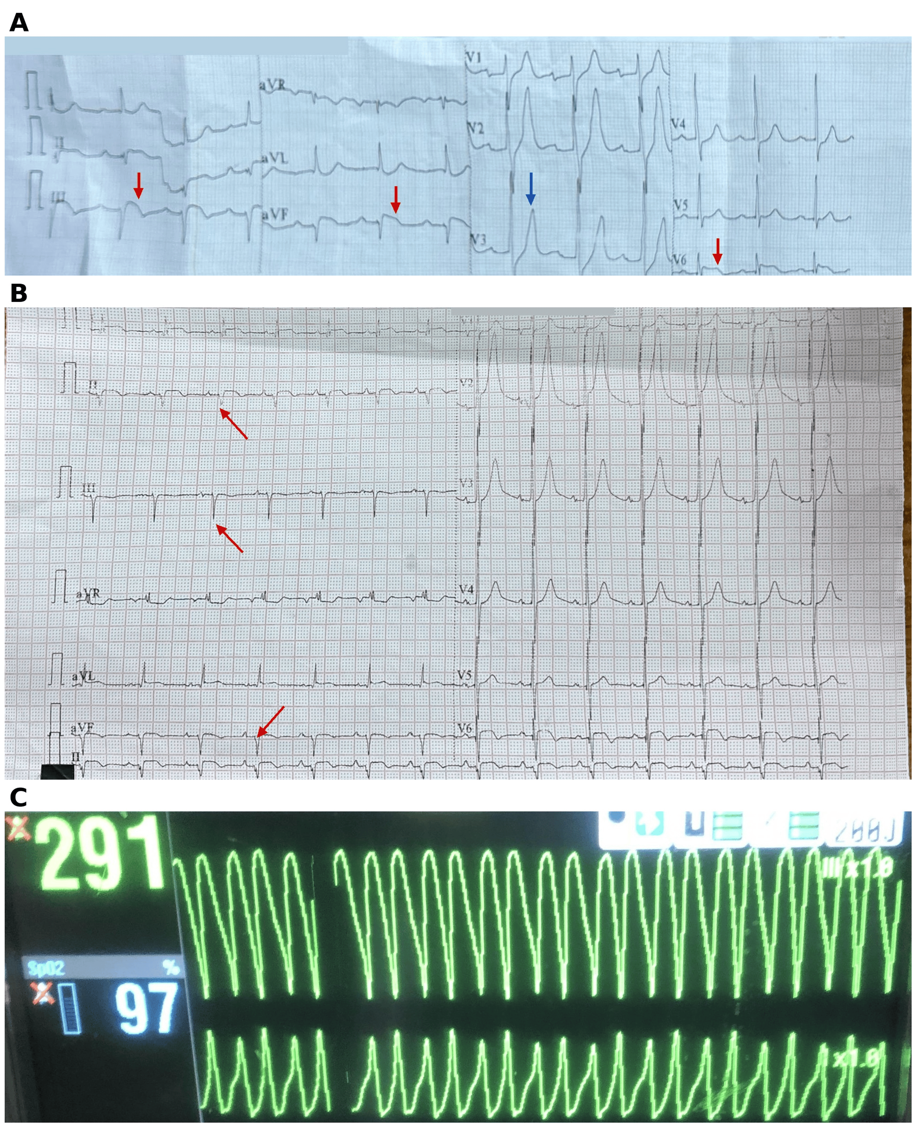
A 53-year-old man with a two- to three-year history of hypertension presented to the emergency department in the evening with chest pain that had begun earlier that day. He had no known drug allergies. Point-of-care testing showed a creatine kinase-MB level above 80 ng/mL, a troponin I level of 9.65 ng/mL, and a B-type natriuretic peptide (BNP) level of 216 pg/mL. The electrocardiogram (ECG) showed ST-segment elevation in leads II, III, aVF, and V6, with ST-segment depression in V1 and tall T waves in V2-V3, consistent with an inferolateral infarction extending to the posterior wall (Figure 1A). By hospital day 2, Q waves had developed in the inferior leads, accompanied by ST-segment elevation and T-wave inversion in leads II, III, aVF, I, aVL, and V6 (Figure 1B).
Blood tests at presentation showed a leukocyte count of 12.49×10³/µL, with 84% neutrophils; hemoglobin of 14.6 g/dL; platelet count of 137×10³/µL; creatinine of 0.96 mg/dL; and potassium of 4.77 mmol/L. Glycated hemoglobin was 9.1%, indicating previously undiagnosed diabetes mellitus. Tests for HIV, hepatitis B surface antigen, and hepatitis C antibodies were negative. Echocardiography before catheterization showed a left ventricular ejection fraction (LVEF) of approximately 50% by visual estimation, hypokinesia of the posterior and lateral walls, and mild mitral regurgitation.
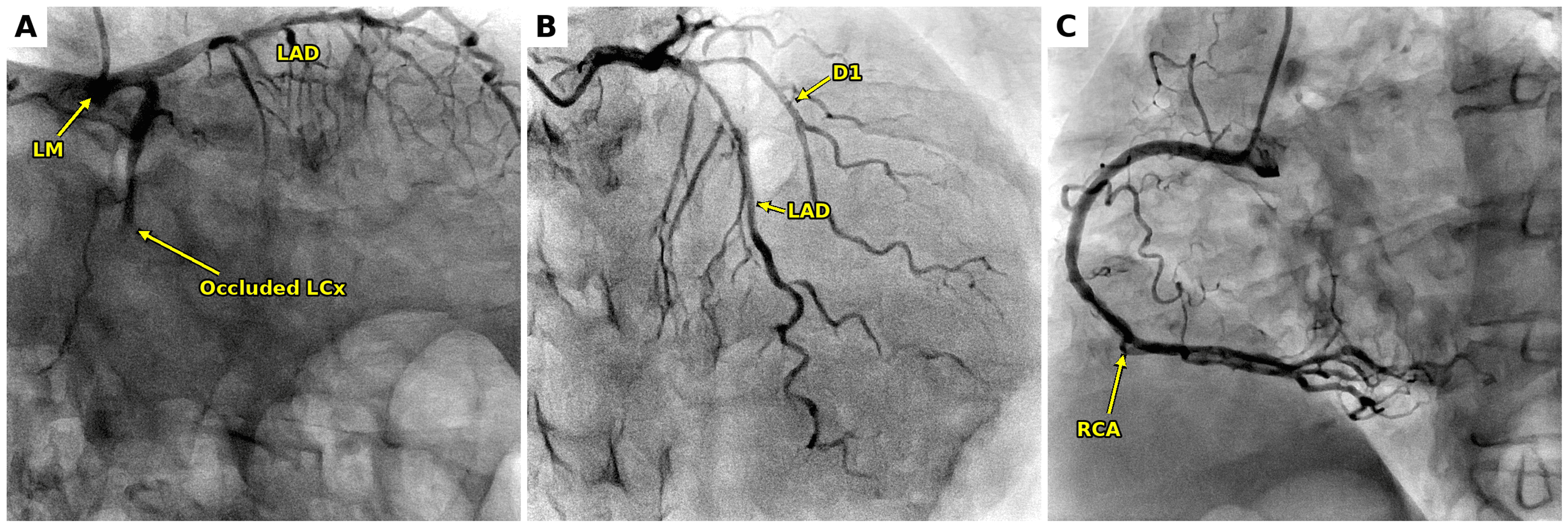
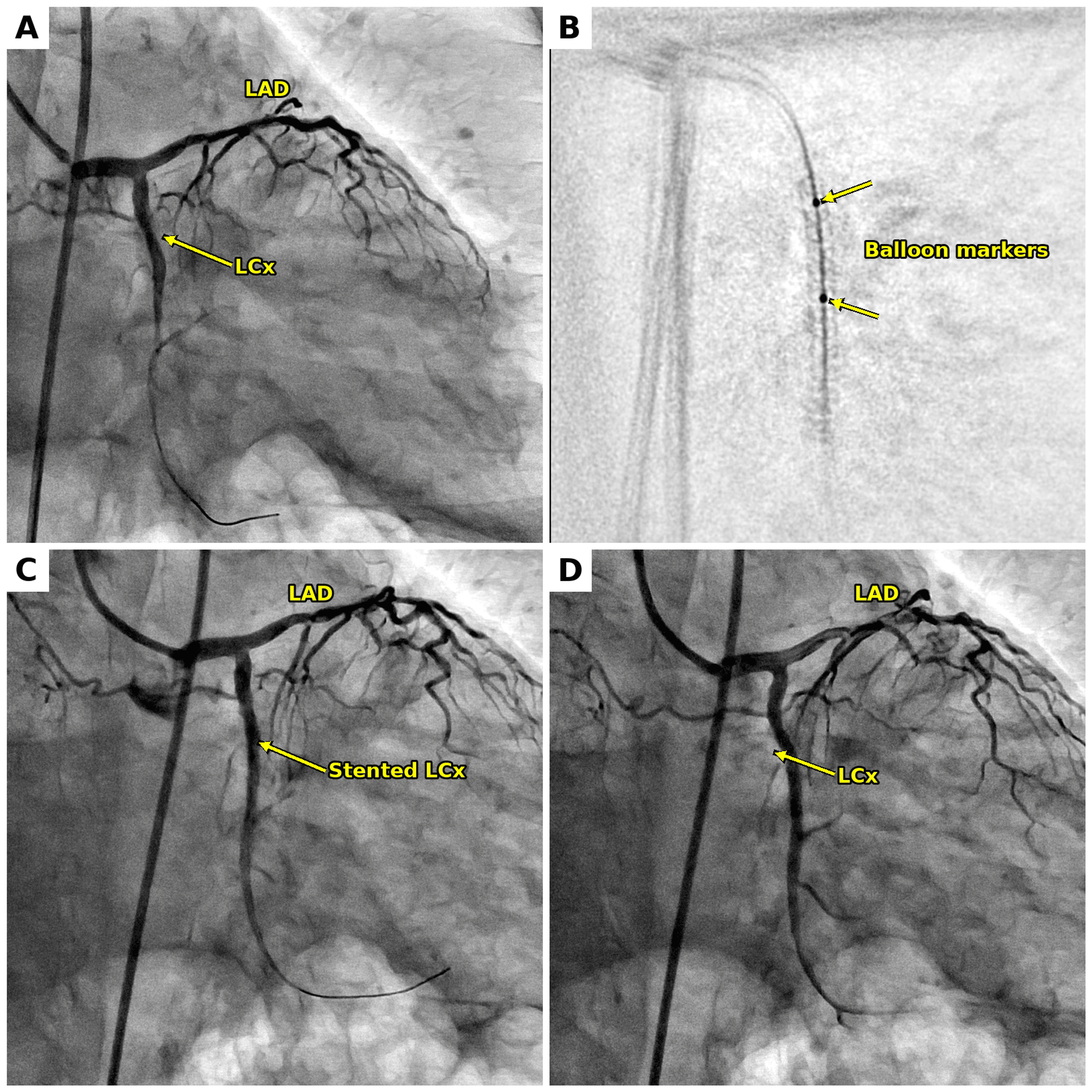
Coronary angiography on the morning of hospital day 2 showed total occlusion of the proximal LCx, judged to be the culprit lesion, with moderate non-culprit disease: 60% stenosis of the mid left anterior descending artery (LAD) and 50%-60% stenosis of the proximal first diagonal branch (Figure 2). Primary PCI of the LCx was performed that afternoon through the right femoral artery under local anesthesia, with 7,000 IU of unfractionated heparin and activated clotting time monitoring. Using a 6-Fr XB 3.5 guide catheter, the occlusion was crossed with a Pilot 50 guidewire (Figure 3A) and predilated with a non-compliant balloon at 12 atm. A 2.5 mm × 28 mm drug-eluting stent was deployed at 14 atm (Figure 3B) and postdilated with a 2.75 mm × 12 mm non-compliant balloon at 16 atm, restoring thrombolysis in myocardial infarction (TIMI) grade 3 flow (Figures 3C, 3D). A tirofiban infusion was administered after the procedure, and treatment with aspirin, ticagrelor, rosuvastatin 40 mg, and metoprolol was initiated. The moderate mid-LAD stenosis was managed medically, with further assessment planned at follow-up.
Leukocytosis with neutrophilia was already present at presentation, before angiography. By the evening of the procedure, the patient had developed a marked systemic inflammatory response: his leukocyte count had risen to 23.51×10³/µL (82% neutrophils), C-reactive protein (CRP) was 272.6 mg/L, and serum sodium had fallen to 129 mmol/L. He also had a cough. Creatinine rose from 0.96 mg/dL to a peak of 4.03 mg/dL on day 4. The AKI occurred after contrast exposure in the setting of a marked systemic inflammatory response and was considered multifactorial [6]. He was treated empirically with meropenem and teicoplanin at renally adjusted doses, intravenous N-acetylcysteine with fluids, oral sodium bicarbonate, and furosemide guided by volume status. Nephrotoxic drugs were avoided. Creatinine fell to 0.84 mg/dL by day 6, whereas CRP declined more slowly, to 216.96 mg/L on day 6 and 142.14 mg/L on day 7. Procalcitonin on day 7 was 2.10 ng/mL. The laboratory course is summarized in Table 1.
On day 6, the patient experienced recurrent syncope caused by sustained monomorphic VT. The defibrillator monitor recorded a regular wide-complex tachycardia at 291/min (Figure 1C), and episodes recurred in rapid succession, meeting the definition of electrical storm. A 12-lead ECG during VT was not available. By then, creatinine had returned to 0.84 mg/dL, but CRP remained 216.96 mg/L, potassium was 3.62 mmol/L, and BNP had risen to 1,020 pg/mL. Treatment consisted of intravenous lidocaine (100 mg bolus, followed by 2 mg/min), repeated 150 mg boluses of intravenous amiodarone followed by an infusion, magnesium sulfate, calcium gluconate, and oral mexiletine 150 mg twice daily, together with repeated synchronized cardioversion. Tirofiban and nicorandil infusions were restarted until repeat angiography could be performed. VT continued to recur at short intervals, so the patient was sedated with midazolam and fentanyl and mechanically ventilated. Repeat angiography on day 7 showed a widely patent LCx stent with TIMI grade 3 flow, excluding stent thrombosis. Between episodes, the ECG showed sinus rhythm with QT prolongation during amiodarone therapy (corrected QT interval up to 489 ms). Electrolytes were monitored closely, and serum magnesium on day 7 was 2.25 mg/dL.
Because antiarrhythmic drugs and repeated cardioversion had failed and the stent was patent, the anesthesia team was consulted for cardiac sympathetic blockade. Immediately before the block, the patient was sedated and mechanically ventilated, and VT continued to recur despite the antiarrhythmic regimen described above. On day 8, a left SGB was performed under ultrasound guidance. A 5 cm Stimuplex needle was advanced just below the carotid artery to the plane superficial to the longus colli muscle, and 6 mL of 1% lidocaine containing 4 mg of dexamethasone was injected. No complications occurred, and the patient remained hemodynamically stable during the procedure. Left-sided Horner syndrome confirmed the effectiveness of the block. VT ceased following the block and did not recur during the remainder of the hospital stay. Amiodarone, mexiletine, and metoprolol were continued. Sedation was withdrawn, and the patient was extubated within 24 hours.
By day 10, the patient was hemodynamically stable (blood pressure, 124/80 mmHg; heart rate, 90/min; oxygen saturation, 98% on room air) and free of arrhythmia, with no further inflammatory or renal complications. He was receiving aspirin, ticagrelor, rosuvastatin, metoprolol 25 mg twice daily, amiodarone 200 mg twice daily, and mexiletine 150 mg twice daily. A secondary-prevention implantable cardioverter-defibrillator (ICD) was advised but could not be implanted during the index admission because of financial constraints; implantation is planned. Electrophysiology review, including consideration of catheter ablation, and reassessment of the LAD stenosis were also planned at follow-up.
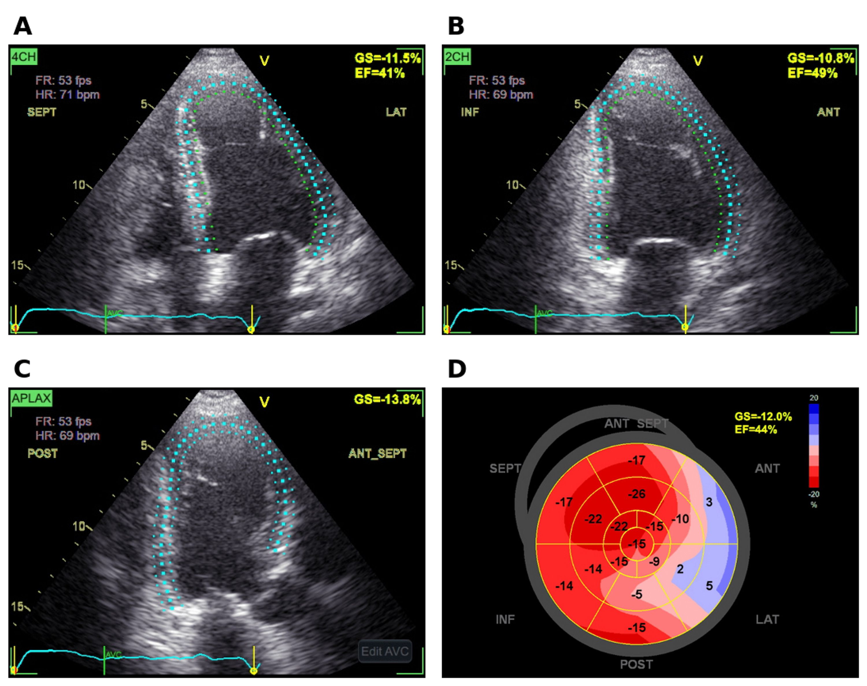
Echocardiography on day 11 showed an LVEF of 40% by the single-plane Simpson method and 44% by speckle tracking, lower than the visual estimate of about 50% before PCI, with a global longitudinal strain of -12.0% (Figure 4). Strain was relatively preserved in the septal, anteroseptal, and inferior segments but was severely reduced or paradoxical (positive) in the anterolateral and basal anterior segments, with abnormality centered on the lateral wall supplied by the LCx. Peak strain dispersion, which reflects heterogeneity in the timing of regional contraction, was 79 ms. Key events are summarized in Table 2.
Discussion
VT storm began on day 6, after the inflammatory and renal complications had peaked. The key question at the bedside was whether the arrhythmia reflected recurrent ischemia due to stent failure, which would have required urgent repeat PCI, or a scar-related mechanism. Several observations favored the latter. The VT was sustained and monomorphic; it began several days after reperfusion rather than within the first 48 hours, when ventricular arrhythmias are usually attributed to acute ischemia, and repeat angiography showed a widely patent stent with TIMI grade 3 flow [1]. Strain imaging also showed regional mechanical dysfunction centered on the lateral wall, the territory of the occluded LCx; impaired strain in the basal anterior segment may reflect overlap with the diagonal territory or the LAD stenosis. A scar-related substrate was therefore considered likely. This remains an inference: a contribution from the moderate non-culprit LAD stenosis could not be completely excluded, and without a 12-lead ECG during VT, cardiac magnetic resonance imaging, or electrophysiological mapping, a specific re-entry circuit could not be demonstrated.
Speckle-tracking echocardiography also provided a measure of arrhythmic risk. Mechanical dispersion, the variation in time to peak strain across segments, was greater in post-infarction patients with documented ventricular arrhythmias than in those without, and in a prospective multicenter study, it predicted arrhythmic events independently of LVEF [7,8]. The peak strain dispersion of 79 ms in this patient is close to the value reported in post-infarction patients with recurrent arrhythmias (85±29 ms) [7]. Strain imaging cannot identify scar or localize a VT circuit in the same way as late gadolinium enhancement imaging or electroanatomical mapping. However, it demonstrates regional mechanical dysfunction corresponding to the infarct territory and may provide supportive evidence of the structural substrate when those modalities are unavailable.
Left SGB interrupts efferent sympathetic outflow to the heart and may reduce sympathetically driven arrhythmia triggers and dispersion of repolarization in the infarct border zone. In an early study of 49 patients with electrical storm after acute MI, sympathetic blockade, including left SGB, was associated with lower mortality than antiarrhythmic therapy guided by advanced cardiac life support [9]. Systematic reviews of published case series found significant reductions in ventricular arrhythmia burden and shocks after SGB [2,10]. In one of these reviews, acute MI was the trigger in 15 of 38 patients [10]. Single-center and multicenter series have since reported fewer arrhythmia episodes and defibrillations after the procedure [3,4]. In a prospective multicenter study of 131 patients with refractory electrical storm, percutaneous SGB reduced treated arrhythmic events by at least half within 12 hours in 92% of patients [5]. These studies report group-level reductions in arrhythmia burden; a single case cannot establish efficacy, and our patient’s response should be interpreted as an individual clinical course rather than evidence that SGB works. The 2022 European Society of Cardiology guidelines recommend catheter ablation for monomorphic VT storm that does not respond to antiarrhythmic drugs and state that autonomic modulation may be considered when ablation is ineffective or not possible (class IIb) [1]. In our ventilated patient, SGB could be performed immediately at the bedside, and Horner syndrome provided a simple clinical indication of a successful block. VT ceased following SGB after conventional antiarrhythmic and electrical therapies had failed. The close temporal association suggests a clinically important contribution from sympathetic blockade, although causality cannot be established because amiodarone, mexiletine, and beta-blockade were continued, the patient remained sedated, and his systemic illness was resolving. Most published series used long-acting agents such as bupivacaine or ropivacaine [4,10]. The persistence of arrhythmia control beyond the expected duration of action of lidocaine in our patient is compatible with interruption of a self-perpetuating cycle of sympathetic activation but may also reflect the cumulative effects of concomitant therapy.
The systemic illness may have lowered the arrhythmic threshold, but we regard it as a possible contributor rather than the cause of the storm. Renal function had recovered by the time VT began, whereas the inflammatory response persisted (CRP, 216.96 mg/L; procalcitonin, 2.10 ng/mL), and potassium was in the low-normal range. Systemic inflammation can increase circulating catecholamine levels; these factors may promote ventricular arrhythmias and complicate antiarrhythmic drug dosing. However, VT ceased on day 8 while inflammatory markers remained elevated (CRP, 142.14 mg/L on day 7), so the inflammatory state alone does not explain the timing of the storm. The inflammatory illness and electrolyte disturbances were therefore managed alongside the arrhythmia rather than regarded as its sole cause.
This report has several limitations. Only a two-lead monitor recording of the VT was available, so the exit site could not be localized from QRS morphology. Neither electrophysiological mapping nor cardiac magnetic resonance imaging was performed; therefore, the proposed scar-related mechanism remains unconfirmed. The case notes record repeated cardioversions but not the exact number of VT episodes or shocks, so the arrhythmia burden before and after each intervention could not be quantified. Because VT ceased while antiarrhythmic drugs, beta-blockade, and sedation were continued, the contribution of SGB cannot be separated from that of concomitant therapy. The cause of the systemic inflammatory response was not established, and the patient was treated empirically with broad-spectrum antibiotics. Finally, follow-up is limited to the index admission. A secondary-prevention ICD has been advised but not yet implanted, and longer-term follow-up, including evaluation for catheter ablation, is needed to determine whether arrhythmia control is durable.
Conclusions
In this patient, refractory monomorphic VT storm after primary PCI ceased following left SGB, administered alongside continued antiarrhythmic therapy. Because this is a single case involving concurrent treatments, the response cannot be attributed to the block alone and should not be generalized. In selected patients with refractory electrical storm, left SGB may be considered an adjunctive treatment and a temporary bridge when conventional therapy has failed, but not a substitute for definitive management, such as catheter ablation or an ICD when indicated. When the culprit stent is patent, and the VT is monomorphic, a scar-related substrate is a reasonable working hypothesis, although residual ischemia due to non-culprit disease should not be dismissed. Strain imaging may provide supportive, but not confirmatory, evidence of such a substrate when cardiac magnetic resonance imaging or electroanatomical mapping is unavailable.
References
- Zeppenfeld K, Tfelt-Hansen J, de Riva M, et al.: 2022 ESC guidelines for the management of patients with ventricular arrhythmias and the prevention of sudden cardiac death. Eur Heart J. 2022, 43:3997-4126. 10.1093/eurheartj/ehac262
- Fudim M, Boortz-Marx R, Ganesh A, et al.: Stellate ganglion blockade for the treatment of refractory ventricular arrhythmias: a systematic review and meta-analysis. J Cardiovasc Electrophysiol. 2017, 28:1460-7. 10.1111/jce.13324
- Fudim M, Qadri YJ, Waldron NH, et al.: Stellate ganglion blockade for the treatment of refractory ventricular arrhythmias. JACC Clin Electrophysiol. 2020, 6:562-71. 10.1016/j.jacep.2019.12.017
- Chouairi F, Rajkumar K, Benak A, et al.: A multicenter study of stellate ganglion block as a temporizing treatment for refractory ventricular arrhythmias. JACC Clin Electrophysiol. 2024, 10:750-8. 10.1016/j.jacep.2023.12.012
- Savastano S, Baldi E, Compagnoni S, et al.: Electrical storm treatment by percutaneous stellate ganglion block: the STAR study. Eur Heart J. 2024, 45:823-33. 10.1093/eurheartj/ehae021
- Mehran R, Dangas GD, Weisbord SD: Contrast-associated acute kidney injury. N Engl J Med. 2019, 380:2146-55. 10.1056/NEJMra1805256
- Haugaa KH, Smedsrud MK, Steen T, et al.: Mechanical dispersion assessed by myocardial strain in patients after myocardial infarction for risk prediction of ventricular arrhythmia. JACC Cardiovasc Imaging. 2010, 3:247-56. 10.1016/j.jcmg.2009.11.012
- Haugaa KH, Grenne BL, Eek CH, et al.: Strain echocardiography improves risk prediction of ventricular arrhythmias after myocardial infarction. JACC Cardiovasc Imaging. 2013, 6:841-50. 10.1016/j.jcmg.2013.03.005
- Nademanee K, Taylor R, Bailey WE, Rieders DE, Kosar EM: Treating electrical storm: sympathetic blockade versus advanced cardiac life support-guided therapy. Circulation. 2000, 102:742-7. 10.1161/01.cir.102.7.742
- Meng L, Tseng CH, Shivkumar K, Ajijola O: Efficacy of stellate ganglion blockade in managing electrical storm: a systematic review. JACC Clin Electrophysiol. 2017, 3:942-9.