

Authors Dhananjay D, Rao R, Chandrashekhar S, et al.
Cureus 18(10): e117945. doi:10.7759/cureus.117945
Abstract
Extensive juvenile-onset recurrent respiratory papillomatosis (JORRP) can turn the paediatric airway into a constantly changing operative field, particularly when disease extends from the larynx into the tracheobronchial tree. Shared airway surgery, the risk of obstruction and the dynamic changes that occur during lesion excision make anaesthetic management especially challenging.
We describe the anaesthetic management of a two-year-old child with extensive laryngotracheobronchial papillomatosis and a long-term tracheostomy after failed decannulation. A staged airway strategy was used, with spontaneous ventilation preserved throughout. At induction, the existing metallic tracheostomy tube was exchanged for a cuffed flexometallic endotracheal tube. After lesion debulking, a significant air leak developed, so the tube was changed to a microlaryngeal surgery (MLS) tube to improve the tracheal seal while maintaining surgical access. For excision of distal airway lesions, the MLS tube was removed, and tubeless oxygenation was provided with transnasal humidified rapid-insufflation ventilatory exchange (THRIVE). The procedure was completed successfully, and the tracheostomy tube was reinserted at the end.
Intraoperative challenges included airway sharing, significant airway leak, and laryngospasm, all of which were managed successfully. Successful management requires anticipation of dynamic airway changes and the ability to transition between different ventilation strategies.
Introduction
Juvenile-onset recurrent respiratory papillomatosis (JORRP) is a chronic disease caused predominantly by human papillomavirus types 6 and 11 and is characterised by recurrent papillomatous lesions of the respiratory tract [1]. The disease may require repeated surgical debulking and, in children with extensive airway involvement, prolonged tracheostomy [2]. Anaesthetic management is particularly challenging because of the shared airway, friability and bleeding potential of the lesions, risk of airway obstruction, and the need to balance adequate oxygenation and ventilation with optimal surgical exposure [3].
In extensive disease, airway requirements may change during the procedure as lesions are progressively removed and the relationship between the airway, tracheal tube, and surgical field changes. When the disease extends into the tracheobronchial tree, lesions lie beyond the tube cuff and cannot be reached without compromising the airway. A pre-existing tracheostomy adds further difficulty, as the stoma produces a leak and complicates tube exchange. Consequently, successful management may require transition between different airway and oxygenation techniques rather than reliance on a single strategy.
We describe the anaesthetic management of a child with extensive laryngotracheobronchial papillomatosis and a long-term tracheostomy, highlighting a staged airway strategy that was modified according to the evolving surgical requirements.
Case Presentation
A two-year-old boy weighing 15 kg and measuring 90 cm, with JORRP, was admitted for evaluation following failed decannulation of a tracheostomy. There was no history of noisy breathing, respiratory distress, or recent respiratory tract infection. Birth and developmental histories were unremarkable. On examination, the child was comfortable at rest with a metallic Jackson tracheostomy tube (5.0-mm internal diameter (ID)) with an inner cannula in situ and demonstrated a good air blast through the tube.
Multiple decannulation attempts had failed; however, the child breathed adequately through the tracheostomy tube. This suggested that the papillomatous lesions above the stoma were causing significant, possibly near-complete, obstruction of the upper airway. On this basis, we decided to place a cuffed flexometallic tube (FMT) through the tracheostomy stoma, to maintain oxygenation and ventilation during the initial excision of these lesions. In view of the extensive papillomatous airway involvement and previous failed decannulation, a potentially unstable airway was anticipated. As an institutional standard practice, a fully equipped difficult airway cart was kept ready.
In the operating room, parental presence was permitted during induction. As the existing metallic tracheostomy tube was incompatible with a universal connector and could not be directly connected to the anaesthesia circuit, anaesthesia was induced via facemask using incremental concentrations of sevoflurane in a 50:50 oxygen-nitrous oxide mixture while maintaining spontaneous ventilation. The metallic tracheostomy tube was not removed before induction. Due to the leak around the uncuffed metallic tracheostomy tube, gas exchange could be maintained despite the tube being in situ. To facilitate mask induction, the tracheostomy tube was temporarily corked, allowing ventilation through the upper airway during induction. The tracheostomy tube was subsequently removed after adequate depth of anaesthesia was achieved, while spontaneous ventilation was maintained. Intravenous access was secured after an adequate depth of anaesthesia had been achieved, and fentanyl 1 µg/kg was administered intravenously.
Spontaneous ventilation was maintained throughout the procedure. The metallic tracheostomy tube was removed and exchanged through the tracheostomy stoma for a 4.0-mm ID cuffed FMT. Although the existing metallic tracheostomy tube had an ID of 5 mm, a 4.0-mm ID cuffed FMT was selected for the exchange, and its insertion was not smooth. In view of the risk of traumatic insertion and creation of a false passage, the tube was not upsized, and the 4.0-mm cuffed FMT was retained after successful placement. Bilateral air entry was confirmed, and the tube was secured to the chest with adhesive dressing. No significant air leak was observed. Dexamethasone 0.2 mg/kg and paracetamol 15 mg/kg were administered for airway oedema prophylaxis and analgesia, respectively.
Bispectral index (BIS) monitoring was applied and maintained between 40 and 60. The vocal cords were topicalised with 10% lignocaine spray, with each puff delivering 10 mg. After induction, the cords were sprayed before the airway was handed over to the surgeon, and a further puff was given every 30 minutes to reduce the risk of perioperative respiratory adverse events. To deliver the drug precisely, a 6 Fr infant feeding tube was attached to the spray nozzle, allowing targeted application to the intended site. This improved efficiency and reduced the dose required. A 20% intravenous lipid emulsion was kept readily available in case of local anaesthetic systemic toxicity.
Following insertion of the suspension laryngoscope, anaesthesia was maintained with propofol target-controlled infusion (TCI) using the Eleveld pharmacokinetic model with effect-site targeting, in an oxygen-air mixture (fraction of inspired oxygen (FiO₂) 0.7-1.0). During maintenance, the propofol effect-site concentration (Cet) ranged from 3.5 to 4.5 µg/mL. Spontaneous ventilation was maintained throughout the procedure and supported with continuous positive airway pressure (CPAP)-pressure support ventilation (PSV) mode of ventilation. Ventilator settings were as follows: mode: CPAP+PSV, FiO2: 0.7-1.0, Psupport: 8 cm H2O, flow trigger: 2.0 L/min, positive end-expiratory pressure (PEEP): 5, minimum respiratory rate (RR): 20/min and inspiratory time (Tinsp): 1.7 seconds. We were able to achieve a tidal volume (TV) ~8 mL/kg with a peak airway pressure ~14 cm H2O. The cuff pressure was around 25 cm of H2O.
Papillomatous lesions were seen in the left aryepiglottic fold, left arytenoid, bilateral true and false vocal cords, and the upper one-third of the trachea (Figure 1), and were excised using coblation. Coblation technology presents virtually no risk of airway fire, even in high-oxygen environments. Coblation (controlled ablation via bipolar radiofrequency plasma) operates at much lower surrounding tissue temperatures than standard electrocautery or lasers. Testing confirms that the device fails to produce flames or ignition when placed in direct contact with flammable materials, such as drapes, prep solutions, or endotracheal tubes, even when flooded with 100% pure oxygen. The coblation device that we used was the Smith & Nephew Coblation II system (Hertfordshire, United Kingdom).
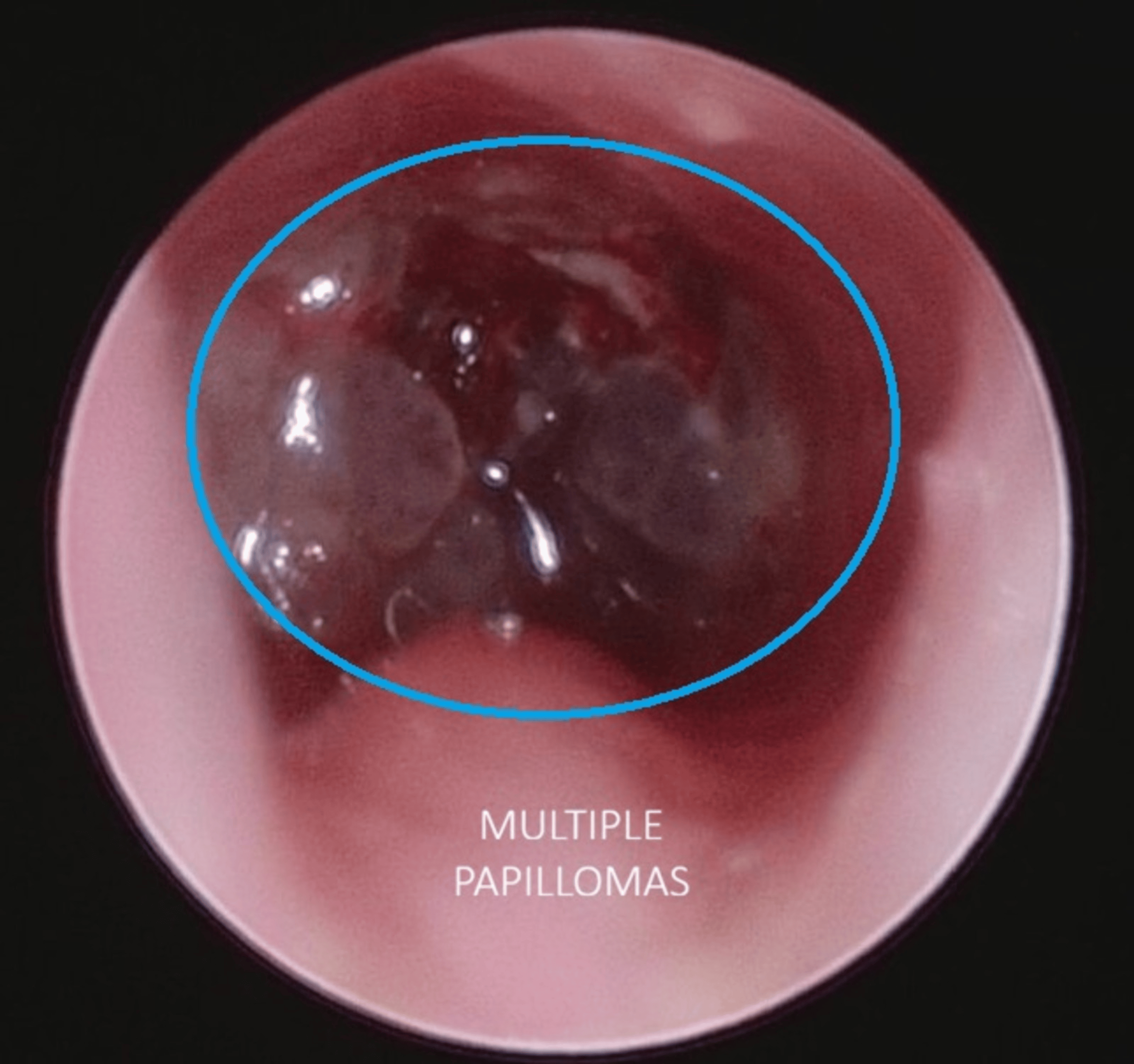
Since there was extensive laryngotracheal papillomatosis (Figure 1), the lesions themselves produced a seal and any possible leak was concealed. Once the infraglottic lesions were excised, the additional seal-effect was lost, and this produced a leak around the cuff of the existing FMT, visualised as bubbling and a drop in the TV to ~4 mL/kg. The FMT was therefore exchanged for a 4.0 mm ID microlaryngeal surgery (MLS) tube. Its larger cuff achieved a more effective tracheal seal, which allowed adequate ventilation to be restored (Figures 2, 3).
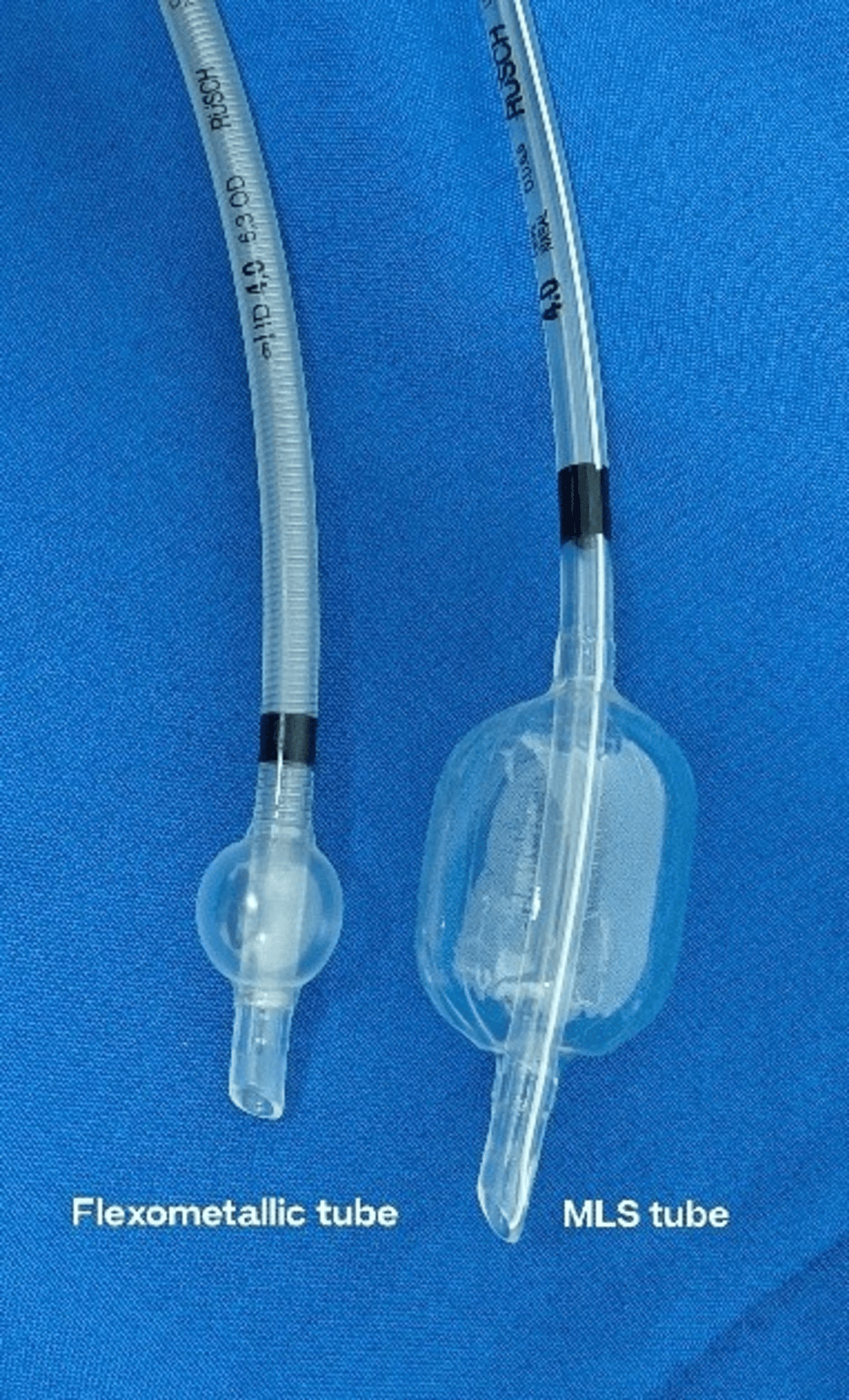
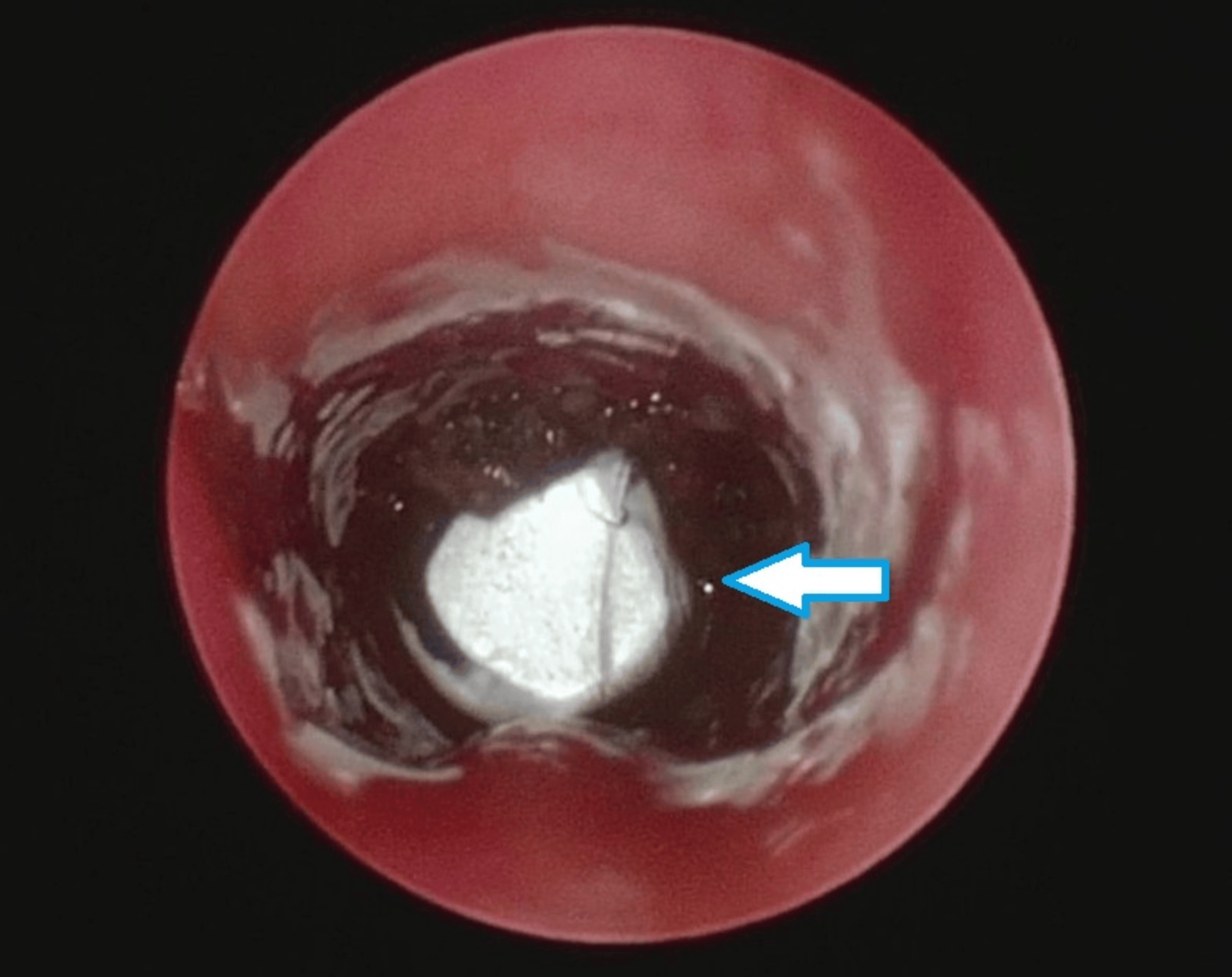
Excision of the lesions located at and beyond the cuff (Figure 4) required unobstructed surgical access. The MLS tube was therefore removed, and the oxygenation strategy was modified. The tracheostomy stoma was sealed with two pieces of gauze secured with a tight elastic adhesive bandage. Tubeless oxygenation was then instituted using transnasal humidified rapid-insufflation ventilatory exchange (THRIVE; Optiflow, Fisher & Paykel, Auckland, New Zealand) at flow rates of 10-20 L/min. THRIVE generates approximately 1 cm H2O of CPAP for every 10 L/min of flow, so these flows corresponded to roughly 1-2 cm H2O. The stomal seal withstood these pressures.
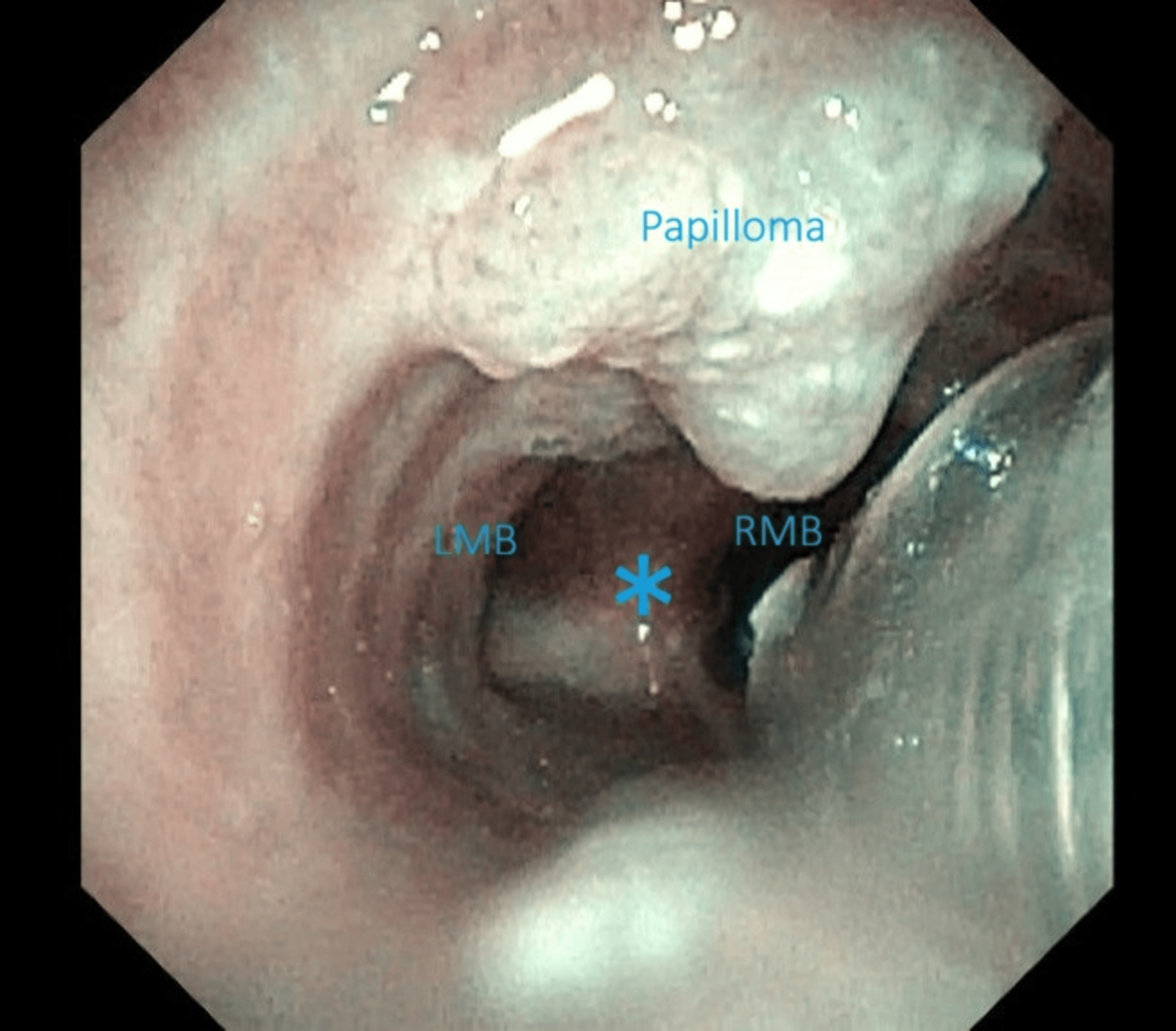
Excision of the lesions located at and beyond the cuff took 38 minutes. Apart from one episode of desaturation, SpO2 remained above 92%. Continuous side-stream capnography was maintained throughout the procedure. However, with an open airway and high-flow insufflation, a reliable quantitative end-tidal CO2 measurement could not be obtained. Transcutaneous CO2 monitoring was not available. SpO2 and visual assessment of chest and abdominal movements served as surrogate indicators of oxygenation and ventilation. No tissue disruption or complications like subcutaneous emphysema were observed.
During the THRIVE phase, one episode of laryngospasm occurred, accompanied by breath-holding and oxygen desaturation to 85%. Vocal cord adduction was visualised on the endoscopic monitor. Oxygenation was continued with THRIVE (FiO2 1.0, flow rate 20 L/min). Surgical stimulation was stopped, and a higher depth of anaesthesia was achieved by increasing the Cet, with BIS-guided titration to prevent apnea. Topical lignocaine was applied to the vocal cords, resulting in prompt resolution of the spasm within a minute. In total, five puffs of topical lignocaine were administered (50 mg, approximately 3.3 mg/kg), which was below the calculated toxic dose of 75 mg (5 mg/kg) for this 15 kg child. Once the laryngospasm was broken, the child continued to breathe spontaneously.
At the end of the 180-minute procedure (Table 1), a 4.0 mm ID uncuffed tracheostomy tube was inserted. The end-tidal carbon dioxide (EtCO2) recorded after connecting to the circle was around 42-44 mmHg. Following resection of the lesions located at and below the cuff, the child’s breathing was supported with CPAP-PSV through the tracheostomy tube, and the EtCO2 fell to 34 mmHg. The total propofol consumption was 362 mg. As the Cet was lowered towards emergence, the child continued to breathe spontaneously and demonstrated appropriate responsiveness and haemodynamic stability.
The child was observed in the post-anaesthesia care unit for an hour to ensure no further peri-operative adverse events. Supplemental oxygen support was delivered using a tracheostomy tube. There were no further airway events, and the child was discharged uneventfully.
Discussion
Anaesthetic management of children with JORRP is complex due to the shared airway, friable lesions, and rapidly changing airway anatomy during surgical debulking [3]. Preservation of spontaneous ventilation during induction is widely recommended in anticipated difficult paediatric airways, as it reduces risk of complete airway obstruction and allows continuous assessment of airway patency [4].
In this case, inhalational induction facilitated safe exchange of the tracheostomy tube to a FMT, while maintaining spontaneous breathing. Progressive lesion removal resulted in a significant air leak, underscoring the importance of anticipating dynamic airway changes and having alternative airway devices available [5]. Use of an MLS tube provided an improved tracheal seal due to its cuff design and larger outer diameter, thereby facilitating effective ventilation during airway surgery [6].
Total intravenous anaesthesia with propofol TCI enabled precise titration of anaesthetic depth guided by BIS monitoring [7]. The Eleveld pharmacokinetic model has been validated across a wide paediatric age range and offers reliable effect-site targeting, making it suitable for complex airway procedures in children [8].
THRIVE has gained increasing acceptance in shared airway surgery by providing effective oxygenation and prolonging safe apnoea time [9]. Its use in paediatric patients has been reported to facilitate airway surgery under spontaneous ventilation and apnoeic conditions [10]. In this case, THRIVE enabled excision of distal airway lesions while maintaining oxygenation during spontaneous respiration. However, risks such as laryngospasm and carbon dioxide accumulation necessitate vigilant monitoring and immediate readiness to deepen anaesthesia or re-establish a definitive airway [9].
Successful management depended on continuous communication between the anaesthesiology and surgical teams and the ability to transition seamlessly between different airway and ventilation strategies, as emphasised in contemporary paediatric airway management recommendations [5].
Conclusions
Extensive laryngotracheobronchial papillomatosis should be approached as a dynamic shared-airway challenge rather than a fixed airway problem. Successful anaesthetic management depends on an adaptable airway strategy, anticipating changes during surgical debulking, maintaining close anaesthesiology-surgical communication and being prepared to transition rapidly between airway and oxygenation techniques to preserve both airway patency and optimal surgical access.
As this report describes a single case, its findings should be interpreted as patient-specific. They do not establish that this management approach is universally applicable, and further cases or controlled studies are needed to confirm its effectiveness
References
- Derkay CS, Wiatrak B: Recurrent respiratory papillomatosis: a review. Laryngoscope. 2008, 118:1236-47. 10.1097/MLG.0b013e31816a7135
- Reeves WC, Ruparelia SS, Swanson KI, Derkay CS, Marcus A, Unger ER: National registry for juvenile-onset recurrent respiratory papillomatosis. Arch Otolaryngol Head Neck Surg. 2003, 129:976-82. 10.1001/archotol.129.9.976
- Bradley J, Lee GS, Peyton J: Anesthesia for shared airway surgery in children. Paediatr Anaesth. 2020, 30:288-95. 10.1111/pan.13815
- Apfelbaum JL, Hagberg CA, Caplan RA, et al.: Practice guidelines for management of the difficult airway: an updated report by the American Society of Anesthesiologists Task Force on Management of the Difficult Airway. Anesthesiology. 2013, 118:251-70. 10.1097/ALN.0b013e31827773b2
- Black AE, Flynn PE, Smith HL, Thomas ML, Wilkinson KA: Development of a guideline for the management of the unanticipated difficult airway in pediatric practice. Paediatr Anaesth. 2015, 25:346-62. 10.1111/pan.12615
- Sarah U, Raju D, Parveen S, Kudpaje A: Transoral laser microlaryngeal surgery with MLT tubes: a retrospective case series. Indian J Otolaryngol Head Neck Surg. 2024, 76:3922-9. 10.1007/s12070-024-04743-3
- Lin YJ, Wang YC, Huang HH, Huang CH, Liao MX, Lin PL: Target-controlled propofol infusion with or without bispectral index monitoring of sedation during advanced gastrointestinal endoscopy. J Gastroenterol Hepatol. 2020, 35:1189-95. 10.1111/jgh.14943
- Eleveld DJ, Colin P, Absalom AR, Struys MM: Pharmacokinetic-pharmacodynamic model for propofol for broad application in anaesthesia and sedation. Br J Anaesth. 2018, 120:942-59. 10.1016/j.bja.2018.01.018
- Riva T, Pedersen TH, Seiler S, Kasper N, Theiler L, Greif R, Kleine-Brueggeney M: Transnasal humidified rapid insufflation ventilatory exchange for oxygenation of children during apnoea: a prospective randomised controlled trial. Br J Anaesth. 2018, 120:592-9. 10.1016/j.bja.2017.12.017
- Harde MJ, Kamble T, Ramchandani P: Apneic anesthesia with THRIVE for pediatric bronchial foreign body removal: a case series. Saudi J Anaesth. 2023, 17:239-41.