

Authors: Kane T M, Russo C M, Chirdon P, et al.
Cureus 18(7): e112650. doi:10.7759/cureus.112650
Abstract
Although giant cell tumors are generally benign, locally aggressive growth can cause profound pathologic tissue destruction and, in some cases, transform into metastatic neoplasms. Here, we present the case of a 37-year-old female with a previously decompressed giant cell tumor of the thoracic spine who presented with acute neurologic symptoms concerning for metastatic transformation requiring a multi-staged surgical resection. The clinical course highlights the potential for late metastatic changes and underscores the importance of long-term surveillance. Our case demonstrates that complex resections performed at historically lower acuity military treatment facilities present additional challenges not seen at higher volume civilian surgical centers. However, it also highlights the importance of engagement among subspecialties across the perioperative period, which led to timely diagnosis and management. This case is noteworthy in that it sparks confidence in the ability of military treatment facilities to successfully collaborate and perform complex, multidisciplinary surgical cases with high fidelity and utmost adherence to the standard of care.
Introduction
Giant cell tumors (GCTs) of the bone comprise roughly 6% of all primary bone tumors. They are classified by the World Health Organization (WHO) as intermediate malignant tumors that rarely exhibit metastatic potential, with a reported incidence of metastatic disease in 2-6% of all confirmed cases [1]. Despite the low likelihood of metastatic disease, GCTs can result in significant life-threatening secondary complications, typically due to local, aggressive expansion causing mass effect on nearby critical anatomical structures such as major vasculature, solid organs, and structures of the peripheral nerves and central nervous system. Aggressive local bone reabsorption is a hallmark feature of the disease. GCTs are made up of numerous multinucleated giant cells undergoing dysregulated growth and significant osteolysis [2]. GCTs are heterogeneous tumors that are composed of several cell types. The two that are most clinically relevant are the giant cells themselves, which are principally responsible for aggressive bone resorption and exhibit many characteristics and properties of osteoclasts; and the stromal cells, which are spindle-like and responsible for the neoplastic properties of GCTs [3]. Giant and healthy osteoclast cells exhibit similarities in behavior, such as their multinucleation and natural ability to resorb bone [4].
Once GCTs are identified via diagnostic imaging studies, they are classified via the Campanacci classification system into grade I (latent phase), grade II (active phase), and grade III (aggressive phase). This grading system is determined by the extent of local damage and aims to quantify the degree of aggressive growth [5]. The Campanacci system is the most frequently utilized system to stage and determine the optimal treatment course and improve outcomes for the patient [6]. The treatment of GCT often requires a delicate balance between making the large surgical excisions needed to address GCTs’ strong propensity to recur, while preserving as much healthy tissue and native structures as possible to preserve patients’ function [7].
Substantial progress has been achieved in the management of these tumors in recent years, notably through pharmacologic treatment alternatives. With the advent of research clarifying the pathophysiology behind GCTs and the role of receptor activator of nuclear factor-κB ligand (RANKL) in the tumor’s highly prolific nature, the United States Food and Drug Administration has approved the use of denosumab, a monoclonal antibody that targets RANKL [8]. Denosumab treatment has been revolutionary for the treatment of GCT; however, the monoclonal antibody treatment introduces diagnostic challenges due to the dramatic depletion and often complete elimination of the characteristic giant cells following therapy. This unique treatment-related alteration of the tumor and subsequent histologic tissues is important to highlight for any clinician who cares for patients with GCTs.
The management of these tumors is complex and multidisciplinary, with large teams comprising orthopedics, neurosurgical, cardiothoracic, plastics, anesthesiology, reconstructive, vascular, and otolaryngology surgical services. A coordinated effort between these services, as well as close collaboration with radiology, pathology, and critical care teams, is tantamount to perioperative success and good postoperative outcomes. The herculean effort involved in coordinating teams is easily overlooked at high-volume, high-acuity quaternary care facilities. This creates a unique challenge when a military or military-affiliated patient is diagnosed with a GCT requiring both medical and surgical management. Compared to large academic hospitals, resources and specialty personnel available within the Military Health System (MHS) are limited and unpredictable; thus, the management of a locally aggressive GCT can stress that system and/or result in delay of the time-sensitive care needed for the patient to result in the best possible outcome. However, with appropriate preoperative planning, interdisciplinary coordination and communication, as well as attention to detail and the combined efforts of flexible and dedicated perioperative teams, these rare and potentially devastating cases can be safely managed within the MHS, with utmost adherence to the standard of care, and with the expectation of strong clinical outcomes. Here, we present a case of the successful management of a local destructive GCT that was treated preoperatively with denosumab resulting in unique pathology at a Military Treatment Facility (MTF) in October 2024.
Case Presentation
A 37-year-old female, with a known history of a GCT of the T2 vertebral body post-surgical decompression and fixation four years prior, presented to the emergency department (ED) with an acute onset of right upper extremity radiculopathy concerning for disease recurrence. The patient had previously undergone a three-year course of monoclonal antibody therapy with denosumab that was subsequently discontinued due to the development of stage 2 medication-induced osteonecrosis of the jaw.
Due to the acute onset of radicular symptoms and the patient’s history of GCT, urgent diagnostic images were obtained (CT and MRI without contrast), which exhibited aggressive tumor growth of the T2 GCT. The tumor now measured 7.1 cm × 4.8 cm, causing cortical bone destruction with increased epidural invasion and mass effect on the upper thoracic spinal cord with evidence of cord compression and edema (Figures 1, 2). Orthopedic spine surgery was considered; it was determined on consultation that her GCT demonstrated aggressive grade 3 expansion with possible metastatic transformation. The patient was counseled on her prognosis and her treatment options.
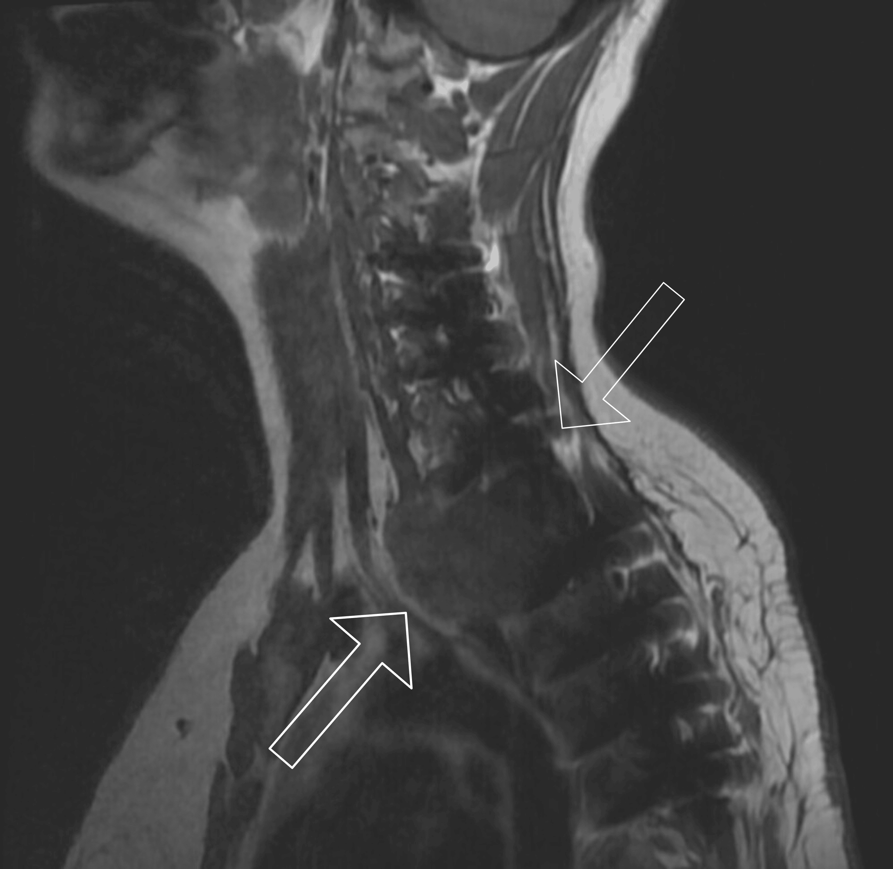
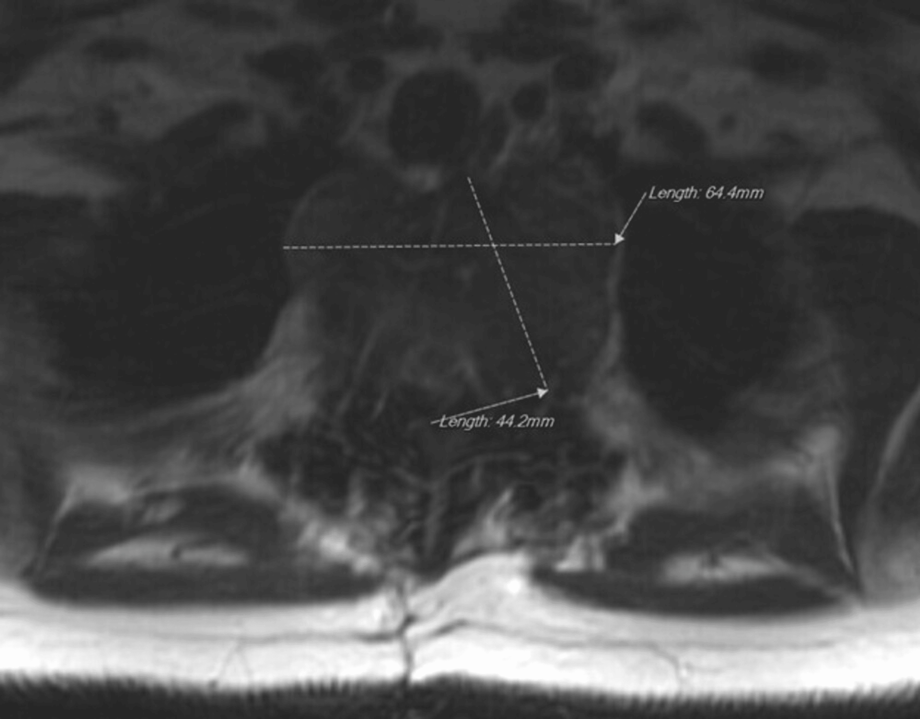
One option was a multi-staged tumor resection, which, due to the tumor location, would require a multidisciplinary surgical team including orthopedic spine, cardiothoracic surgery, head and neck (ENT) surgery, plastic and reconstruction surgery, cardiothoracic anesthesiology, hematology and oncology, acute pain management, and critical care medicine. Following multiple multidisciplinary meetings among the aforementioned teams, the patient, and her family members, the decision was made to proceed with the staged operations, starting with the posterior approach, to be followed four days later by the anterior approach.
Stage 1: Posterior approach
The patient was transported to the operating room, and pre-induction, a right radial arterial line and a left femoral central venous catheter were placed without complications. The patient tolerated induction of general anesthesia without any significant hemodynamic changes and was successfully intubated with a 7.0 standard endotracheal tube (ETT). Confirmation of ETT placement was confirmed via auscultation, end-tidal capnography, and flexible bronchoscopy. Due to the tumor’s proximity to critical structures of the airway (trachea and left main stem bronchus), a bronchial blocker was then deployed via bronchoscopy into the left main stem bronchus and inflated to confirm adequate seal, then subsequently deflated for utilization later in the operation if one-lung ventilation was required. The patient was then put into the prone position, and the neuromonitoring technician established an adequate baseline and signal strength for somatosensory evoked potentials (SSEP) and motor evoked potentials (MEP) to be monitored for the duration of the operation.
A posterior hip incision was made over the patient’s prior surgical scar, and all the preexisting surgical hardware was subperiosteally exposed from C4 to T6 and removed. A C7-T4 laminectomy was performed, and the tumor was notably encountered on the posterior elements of T2. Upon completion of the laminectomy and the spinal cord fully visualized, it was notably displaced dorsally. The tumor was subsequently debulked and excised from a posterior to anterior fashion, and rods were placed from the side-to-side connector to T6 using a Z-rod. The rods were secured with screws to ensure complete spine stabilization in the postoperative period in anticipation of the second stage (anterior approach) four days later.
Due to the length of the operation, the intraoperative estimated blood loss of 6,500 mL (with an intraoperative hemoglobin nadir of 5.9 g/dL), and concern for the possible development of coagulopathy, the patient received a balanced transfusion of blood products that included 12 units of packed red blood cells, 12 units of fresh frozen plasma, and 2 units of platelets. Upon surgical closure and completion of the operation, the patient remained intubated and went from the operating room to radiology for a postoperative CT scan and then was transported in stable condition to the surgical intensive care unit (SICU), where she was extubated without issue, moving all extremities, and at neurologic baseline.
Initial postoperative course
Overnight, the patient remained hemodynamically stable and required no additional blood products or vasoactive medications to maintain a mean arterial pressure greater than 60 mmHg. She did, however, have multiple desaturation events and complained of progressing dyspnea and pleurisy. By the morning, she required 8 L of oxygen via a nasal cannula to maintain SpO2 saturations above 90%.
Due to the length of the procedure and anatomical location, as well as the massive fluid shifts expected from significant surgical blood loss and subsequent blood product transfusions, concerns for atelectasis, pneumothorax, pulmonary embolism, and infection were on the differential diagnosis. A CT pulmonary angiogram was performed on postoperative day one, which showed bilateral pulmonary embolisms with possible early evidence of right heart strain.
Due to the location of the operation and concerns about the formation of a potential epidural hematoma, the patient was deemed not a good candidate for chemical anticoagulation; hence, the vascular surgery service was consulted for inferior vena cava filter placement. This was placed the following day under fluoroscopic guidance without complication. The patient recovered over the following days in preparation for the second stage of her operation.
Stage 2: Anterior approach
Four days following the posterior approach, the patient was transported back to the operating room for the second stage of the surgery. The patient underwent induction of general anesthesia and was intubated with a 37 F left-sided double-lumen tube (DLT) without any complications. Neuromonitoring leads were placed, and adequate SSEP/MEP baselines and signals were established.
The anterior approach started with a large apron incision and dissection. A platysmal flap was then raised for optimal surgical visualization. A left-sided clavicle osteotomy was performed on both sides of the clavicular insertion of the sternocleidomastoid (SCM) to allow the SCM to be elevated, for the subclavian vessels and nerves to be visualized, and for safe dissection without complication. The carotid sheaths and strap muscles were dissected to allow visualization of the anterior spine. The brachial plexuses and recurrent laryngeal nerves were then identified and dissected bilaterally to ensure protection throughout the remainder of the operation. The dissection was then extended posteriorly until the inferior trunks of the brachial plexus were visualized. At this time, with partial tumor visualization, it was noted that the T1 and C8 nerve roots were draped over the tumor, and the left subclavian and vertebral arteries coursed over the tumor. A level five neck dissection was completed, and all neurovascular structures, including the brachial plexus, were dissected free from the tumor. The esophagus was safely dissected and mobilized. A left-chest wall midaxillary incision was made for left-sided mass visualization, followed by a right-sided thoracotomy for visualization of the right side of the mass. With the tumor fully dissected and visualized, it was divided into two lobes, and the lobes were subsequently removed without complication.
With the tumor completely removed and hemostasis obtained, the focus turned to the reconstruction phase of the operation. A vascular harvest of the left fibula as the C7 and T3 vertebral bodies were prepared for a press-fit graft. A composite graft was then placed spanning C7-T3 with excellent press-fit and intrinsic stability. The anterior cervical plate was contoured and placed from C6-T3 and was fixed proximally and distally with 16 mm titanium screws through the plate. Upon completion of the composite graft, the vascular graft anastomosis was achieved, and surgical closure was carefully performed without complication.
During this period, the patient exhibited significant challenges with oxygenation, which progressively worsened 10 hours into the case. Furthermore, pulmonary pressures gradually increased, and her tidal volume (TV) progressively decreased in an inverse relation to the length of the procedure. Ventilator settings were adjusted to address tissue oxygenation concerns, including increasing peak end-expiratory pressure and a reduction of TV with an increase in respiratory rate. Unfortunately, there was little improvement. Flexible bronchoscopy was performed via CT anesthesia to confirm placement of the DLT and to provide intratracheal suction. Additionally, arterial blood gases were trended in a serial fashion and were concerning for a PaO2 nadir of 68 mmHg on 100% FiO2.
Upon conclusion of the reconstruction phase of the operation, in light of the patient decompensating from a pulmonary perspective, she was transferred intubated and sedated to the SICU.
Pathology
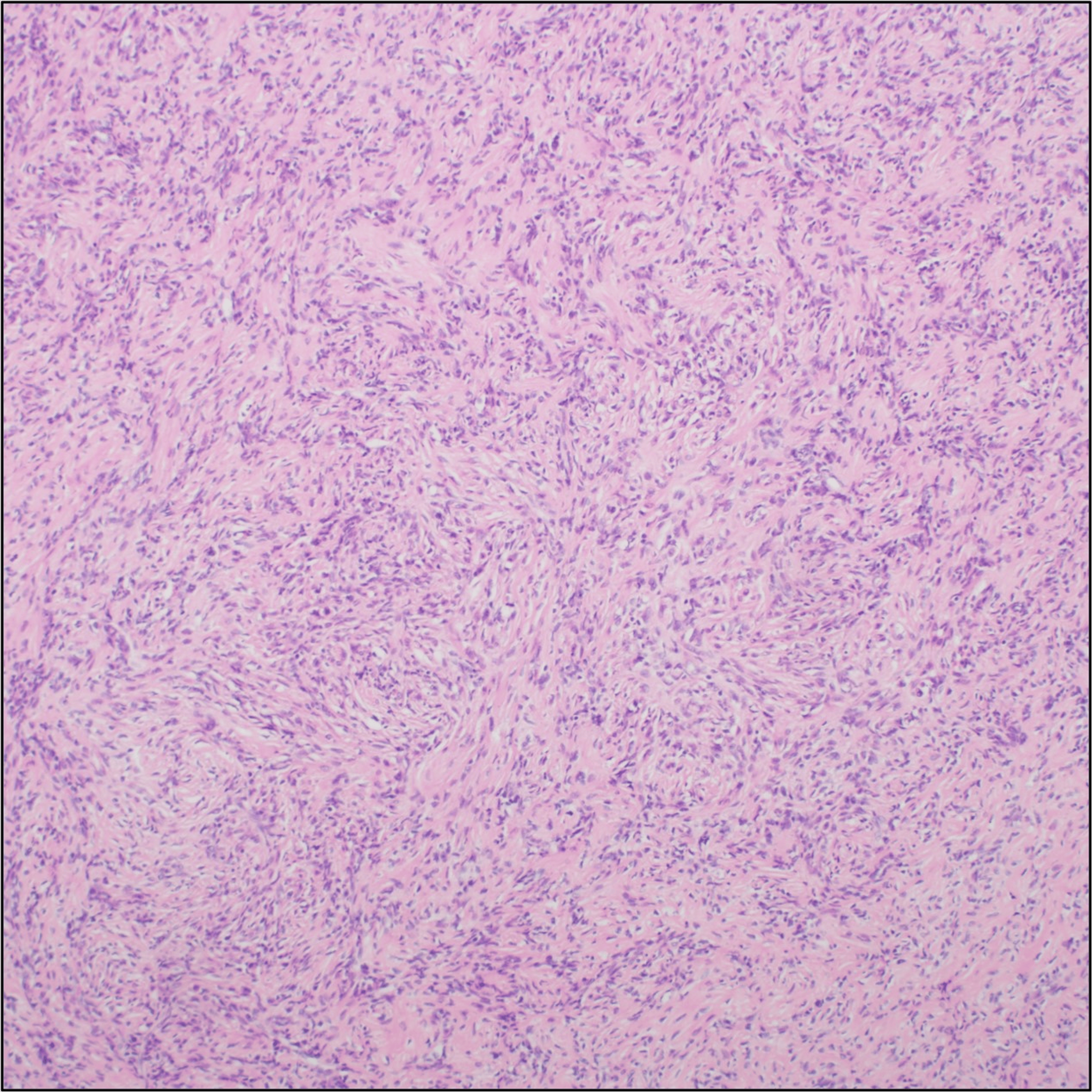
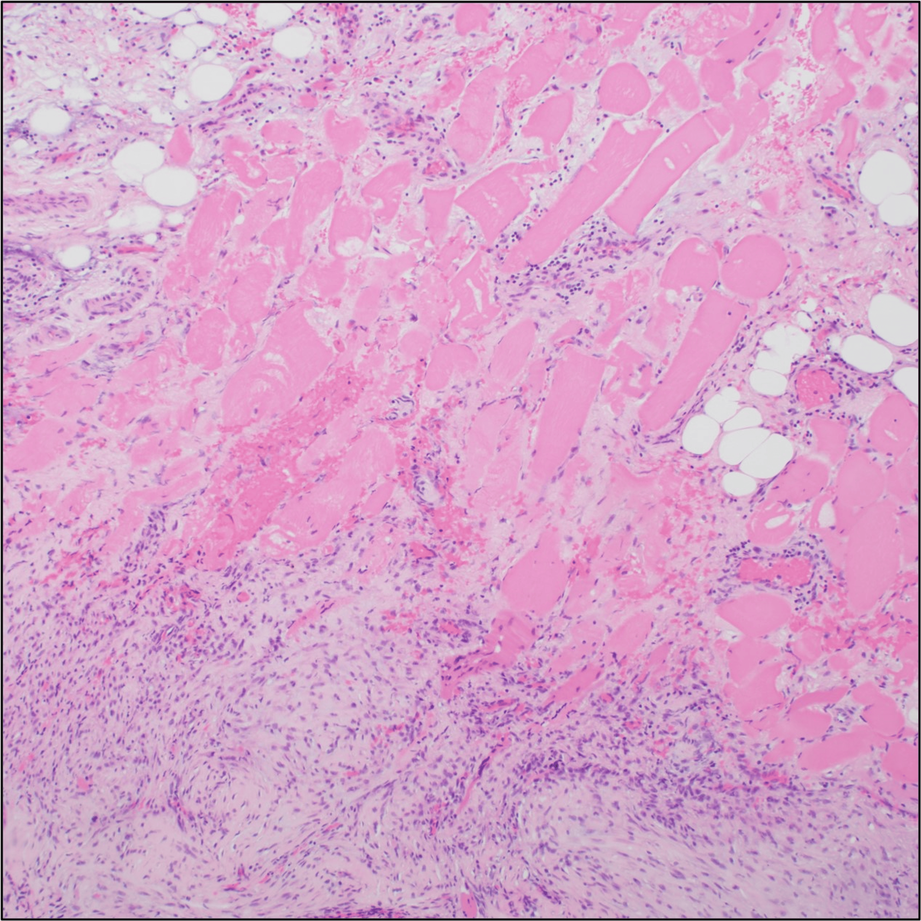
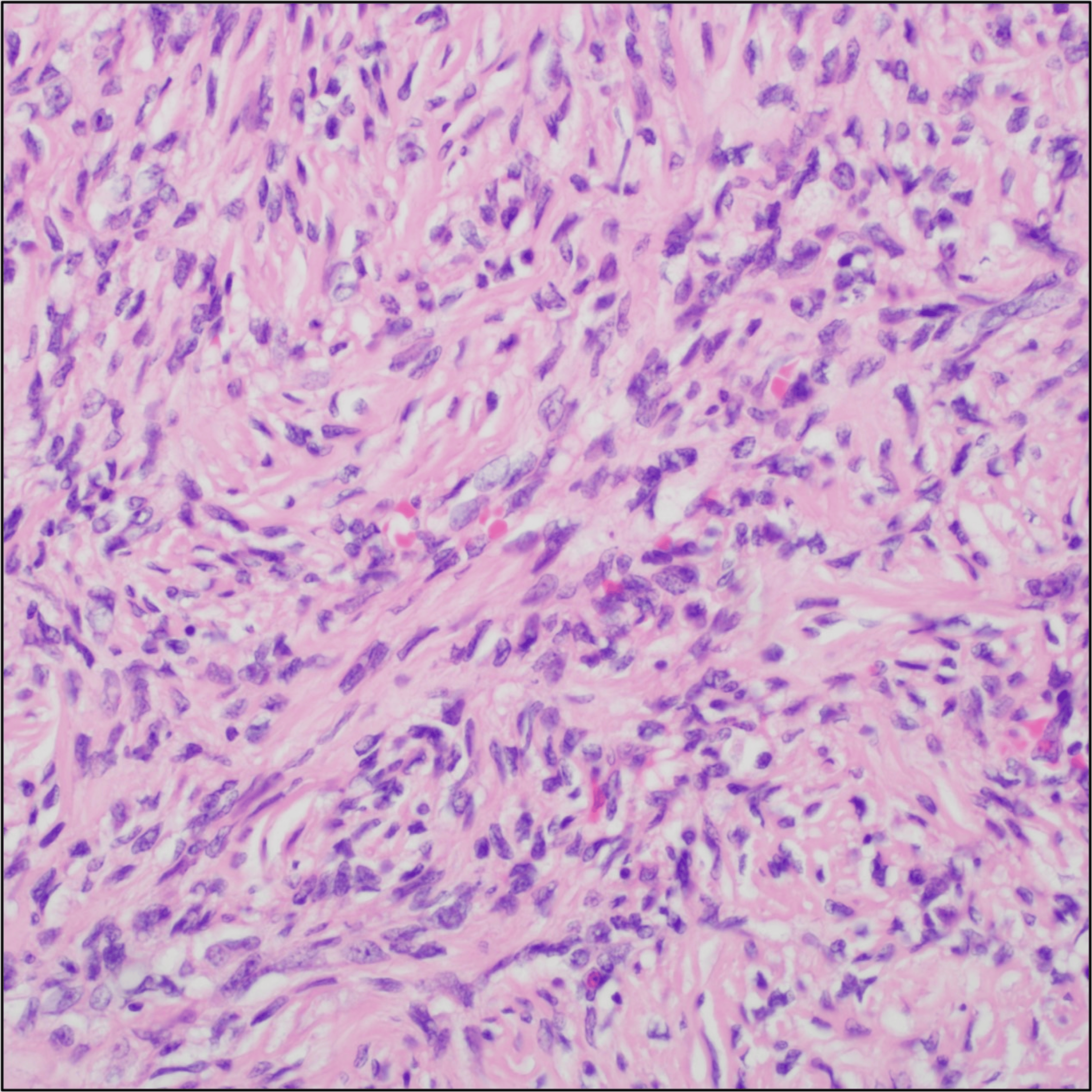
Histology showed richly vascular, nodular proliferation of spindle cells growing in short, storiform fascicles with areas of chondroid metaplasia and new bone formation. There were areas where the lesion broke through or obliterated the native bone cortex and grew out into the surrounding fibroadipose tissue and skeletal muscle. Immunohistochemistry was performed for H3G34W (H3.3 p.Gly34Trp), which is the hallmark diagnostic marker for GCTs of the bone, with adequate controls, and was diffusely positive. Amplification of MDM2 was not detected by fluorescence in situ hybridization. The uniform reactivity for H3G34W supported the diagnosis of GCT of the bone. Furthermore, the patient’s tumor was notably without any giant cells. This is consistent with the history of denosumab treatment and highlights a unique and clinically important aspect of the case (Figures 3–5).
Discussion
The diagnosis of GCT can be challenging to identify on time, particularly if the tumor is located within the spine. With no obvious gross deformity presenting itself on physical examination, compared with, for example, a tumor arising from an extremity such as a femur, tibia, or radius, GCTs of the spine are often identified as incidental radiographic findings or discovered upon the presentation of acute neurological deficits and pain [9].
The correct diagnosis of a GCT is only the first of many hurdles in a long process of perioperative management, multidisciplinary consultation, surgical and anesthetic planning, and recovery and rehabilitation after a multi-staged operation. In addition to the perioperative challenges and surgical management of GCTs, their elevated rate and propensity for recurrence make the postoperative follow-up phase of care key for the patients’ overall successful outcome.
Aligning these numerous medical specialties, as well as the ancillary staff needed to perform such a multi-staged resection of a large GCT of the thoracic spine, would be challenging enough at a high-volume, high-acuity major tertiary medical center, which would typically have access to an abundance of resources and specialists who routinely care for cases of this magnitude regularly. Performing this complex series of procedures and postoperative care at an MTF is far more of a challenge. MTFs are littered with unique challenges that can make the decision to attempt complicated surgical cases difficult for military surgeons.
It is well known and described in the literature that consistent procedural volume is widely inconsistent among MTFs and varies significantly based on location [10]. Furthermore, the MHS is constantly balancing its dual mission of providing comprehensive medical services for covered beneficiaries and ensuring active-duty medical personnel are ready for deployment [11]. Of these two often competing interests, deployment readiness takes precedence. This prioritization and investment in readiness have resulted in many high-value surgeries within the MHS being covered under “purchased care” or referred to civilian facilities.
A comprehensive retrospective analysis of surgical volumes within all MTFs was conducted in 2021 that examined 292,411 high-value surgical cases from 2005 to 2019. High-value cases were identified using a knowledge, skill, and attitude (KSA) metric that was developed by the Department of Defense (DoD) to evaluate the transferable skills incorporated into a given surgery or medical procedure that are considered essential or highly relevant for surgeons to maintain deployment readiness [11]. The surgical cases identified were pancreatectomies, hepatectomies, esophagectomies, colectomies, coronary artery bypass grafts, abdominal aortic aneurysm repairs, and carotid endarterectomies. More than 90% of all examined cases were referred to the civilian sector for “purchased care,” with the proportion of cases referred increasing over the study period.
Presumably due to the high medical screening standards for active-duty service members, the age of the active-duty population, and an overall lack of pre-existing comorbidities, there is a relatively low volume of surgical pathology at most MTFs, which therefore have relatively few large multi-stage complex surgeries. Most cases that do arise are transferred to a civilian medical center; there is little left at the MTF for active-duty medical personnel to manage in terms of complex perioperative surgical cases. Despite this lack of surgical volume and case complexity, however, multiple studies have shown that surgeries performed at MTFs are associated with fewer complications [12].
Additional concerns regarding surgical workload and its downstream effects on deployment readiness have been thoroughly studied. In a period of just four years from 2015 to 2019, the number of general surgery procedures generating KSA in military hospitals decreased by 19%. This corresponded to a notable decrease in the number of active-duty general surgeons meeting the KSA metrics for deployment readiness, with this percentage falling from 17% in 2015 to less than 10% in 2019 [13]. The intersection of cost, patient safety, deployment readiness, and high-value active-duty medical personnel retention is a balancing act with countless variables and consequences not only for beneficiaries of the TRICARE healthcare system for military personnel but also for the future conflict preparedness of the United States.
In an effort to maintain essential surgical skills and capabilities, the US DoD has made significant strides in partnering with established high-volume civilian surgical centers. It has also ensured the exposure of active-duty DoD medical assets to specific pathology and case complexity at civilian centers [14]. However, the placement of select numbers of the DoD’s most highly trained medical specialists outside MTFs has resulted in several unintended downstream consequences, namely, a reduced ability to provide complex surgical care within MTFs. Considering the nuanced challenges related to managing complex high-risk perioperative surgical patients, significant additional effort and collaboration are required for these procedures within MTFs to ensure the best possible outcome [15].
Multidisciplinary planning in healthcare is a critically important and emerging field of research that examines and evaluates patient outcomes, complications, costs, system optimization, resource allocation, and utilization. Within the perioperative setting, it has been shown that the use of multidisciplinary surgical teams not only results in superior patient outcomes but is cost-effective for both healthcare facilities and patients [16]. Furthermore, multiple systematic reviews highlight the importance of interdisciplinary collaboration across various specialties and its positive effects on medical and graduate medical education for trainees, regardless of their level of training [17-19]. There is little to no evidence in the current literature that multidisciplinary collaboration results in any additional negative outcomes for hospital systems, patient outcomes, or medical education and training.
As the trend toward specialization and sub-specialization continues among physicians, the collaboration required across different specialties is also increasing. Our case highlights this reality using a complex multi-staged resection of a thoracic GCT, demonstrating the remarkable effort, communication, and teamwork required within an MTF that has historically experienced very few high-risk cases. Adding to the diagnostic complexity of this case was the patient being previously treated with denosumab in an effort to reduce the tumor size before resection. Denosumab therapy often results in histopathologic changes, namely, the reduction or, as in this case, complete elimination of the tumor’s characteristic osteoclastic giant cells. The complex diagnosis and the multidisciplinary collaboration in the perioperative stage, followed by a massive multi-staged surgery involving numerous specialties without significant complications, serve as a testament to the DoD’s capabilities, deployment readiness, and heightened ability to collaborate and perform such procedures despite the lower case volumes and resource limitations faced by many MTFs.
Conclusions
GCTs of the bone are rare and represent a minority of all primary bone tumors. Although the vast majority of GCTs are benign, on rare occasions, metastatic disease has been reported in the literature. Due to GCTs’ unregulated growth and frequent mass effect, when they do arise from the spine, they require multidisciplinary perioperative collaboration between numerous medical specialties to successfully treat the patient. Furthermore, due to the significantly elevated risk of tumor recurrence, a delicate balance must be preserved between the extensive surgical resections needed to reduce recurrence while optimizing the preservation of healthy tissue. Performing an adequate resection of large GCTs involving the spine is virtually always performed with multiple collaborating surgical teams and typically requires multiple staged operations. Due to the complex nature and involvement of numerous medical and surgical specialties that may at times have competing interests, it is paramount to maximize communication and peer engagement between the multidisciplinary teams for optimum perioperative surgical planning. This mentality, however, must extend beyond the operating room and continue throughout the patient’s recovery and long-term follow-up, as the true success of such a major oncological operation is predicated on the patient’s long-term outcome and functional status postoperatively. Performing large multi-staged oncologic tumor resections involving the spine at MTFs can also introduce additional challenges that would largely be mitigated at higher volume civilian surgical centers that routinely perform these complex operations. This case report highlights the diagnostic and multidisciplinary perioperative surgical challenges involved in managing GCTs of the thoracic spine at an MTF where such complex operations are infrequently performed.
References
- Tsukamoto S, Mavrogenis AF, Kido A, Errani C: Current concepts in the treatment of giant cell tumors of bone. Cancers (Basel). 2021, 13:3647. 10.3390/cancers13153647
- Gupta R, Seethalakshmi V, Jambhekar NA, Prabhudesai S, Merchant N, Puri A, Agarwal M: Clinicopathologic profile of 470 giant cell tumors of bone from a cancer hospital in western India. Ann Diagn Pathol. 2008, 12:239-48. 10.1016/j.anndiagpath.2007.09.002
- Cowan RW, Singh G: Giant cell tumor of bone: a basic science perspective. Bone. 2013, 52:238-46. 10.1016/j.bone.2012.10.002
- Vignery A: Osteoclasts and giant cells: macrophage-macrophage fusion mechanism. Int J Exp Pathol. 2000, 81:291-304. 10.1111/j.1365-2613.2000.00164.x
- Campanacci M, Baldini N, Boriani S, Sudanese A: Giant-cell tumor of bone. J Bone Joint Surg Am. 1987, 69:106-14.
- Abbasi AN, Qamar J, Habib A, Ebad Ali SM, Ahmed S, Khan MW: Unraveling the mystery: a comprehensive review of multidisciplinary strategies for managing giant cell tumor of the bone. J Orthop Rep. 2025, 4:100384. 10.1016/j.jorep.2024.100384
- Puri A, Agarwal M: Treatment of giant cell tumor of bone: current concepts. Indian J Orthop. 2007, 41:101-8. 10.4103/0019-5413.32039
- Singh AS, Chawla NS, Chawla SP: Giant-cell tumor of bone: treatment options and role of denosumab. Biologics. 2015, 9:69-74. 10.2147/BTT.S57359
- Leggett AR, Berg AR, Hullinger H, Benevenia JB: Diagnosis and treatment of lumbar giant cell tumor of the spine: update on current management strategies. Diagnostics (Basel). 2022, 12:857. 10.3390/diagnostics12040857
- Carius BM, April MD, Schauer SG: Procedural volume within military treatment facilities-implications for a ready medical force. Mil Med. 2020, 185:e977-81. 10.1093/milmed/usaa001
- Haag A, Cone EB, Wun J, et al.: Trends in surgical volume in the military health system-a potential threat to mission readiness. Mil Med. 2021, 186:646-50. 10.1093/milmed/usaa543
- Haag A, Hosein S, Lyon S, et al.: Outcomes for arthroplasties in military health: a retrospective analysis of direct versus purchased care. Mil Med. 2023, 188:45-51. 10.1093/milmed/usac441
- Dalton MK, Remick KN, Mathias M, Trinh QD, Cooper Z, Elster EA, Weissman JS: Analysis of surgical volume in military medical treatment facilities and clinical combat readiness of US military surgeons. JAMA Surg. 2022, 157:43-50. 10.1001/jamasurg.2021.5331
- Schwab CW: Winds of war: enhancing civilian and military partnerships to assure readiness: white paper. J Am Coll Surg. 2015, 221:235-54. 10.1016/j.jamcollsurg.2015.04.014
- Leeftink AG, Bikker IA, Vliegen IM, Boucherie RJ: Multi-disciplinary planning in health care: a review. Health Syst (Basingstoke). 2018, 9:95-118. 10.1080/20476965.2018.1436909
- Davis MJ, Luu BC, Raj S, Abu-Ghname A, Buchanan EP: Multidisciplinary care in surgery: are team-based interventions cost-effective?. Surgeon. 2021, 19:49-60. 10.1016/j.surge.2020.02.005
- Baker VO, Cuzzola R, Knox C, et al.: Teamwork education improves trauma team performance in undergraduate health professional students. J Educ Eval Health Prof. 2015, 12:36. 10.3352/jeehp.2015.12.36
- Warren MB, Hutchison EP, DeHaan J, Krause AJ, Murphy MP, Velez ML: Residents teaching residents: results of an interdisciplinary educational endeavor. Acad Psychiatry. 2018, 42:473-6. 10.1007/s40596-017-0864-4
- Phillips CB, Hall S, Irving M: Impact of interprofessional education about psychological and medical comorbidities on practitioners’ knowledge and collaborative practice: mixed method evaluation of a national program. BMC Health Serv Res. 2016, 16:465. 10.1186/s12913-016-1720-z